Comparison of end-to-end Gambee anastomosis and triangular anastomosis for esophagogastric anastomosis after subtotal esophagectomy in patients With Thoracic Esophageal Cancer: A Prospective Randomized Controlled Trial
Published in Annals of Thoracic Surgery
Naoaki Maeda1
Naoaki Maeda- 1 Department of Gastroenterological Surgery, Okayama University, Graduate School of Medicine, Dentistry and Pharmaceutical Sciences, Okayama, Japan
Kazuhiro Noma1 ✉
Kazuhiro NomaCorresponding author- 1 Department of Gastroenterological Surgery, Okayama University, Graduate School of Medicine, Dentistry and Pharmaceutical Sciences, Okayama, Japan
Toshiharu Mitsuhashi2
Toshiharu Mitsuhashi- 2 Center for Innovative Clinical Medicine, Okayama University Hospital, Okayama, Japan
Tomoyoshi Kunitomo1
Tomoyoshi Kunitomo- 1 Department of Gastroenterological Surgery, Okayama University, Graduate School of Medicine, Dentistry and Pharmaceutical Sciences, Okayama, Japan
Masashi Hashimoto1
Masashi Hashimoto- 1 Department of Gastroenterological Surgery, Okayama University, Graduate School of Medicine, Dentistry and Pharmaceutical Sciences, Okayama, Japan
Satoru Kikuchi1
Satoru Kikuchi- 1 Department of Gastroenterological Surgery, Okayama University, Graduate School of Medicine, Dentistry and Pharmaceutical Sciences, Okayama, Japan
Toshiaki Ohara1,3
Toshiaki Ohara- 1 Department of Gastroenterological Surgery, Okayama University, Graduate School of Medicine, Dentistry and Pharmaceutical Sciences, Okayama, Japan
- 3 Department of Pathology and Experimental Medicine, Okayama University Graduate School of Medicine, Density, and Pharmaceutical Science, Okayama, Japan
Shunsuke Tanabe1
Shunsuke Tanabe- 1 Department of Gastroenterological Surgery, Okayama University, Graduate School of Medicine, Dentistry and Pharmaceutical Sciences, Okayama, Japan
Yasuhiro Shirakawa4
Yasuhiro Shirakawa- 4 Department of Gastroenterological Surgery, Okayama University, Graduate School of Medicine, Dentistry and Pharmaceutical Sciences, Okayama, Japan; Department of Surgery, Hiroshima City Hiroshima Citizens Hospital, Hiroshima, Japan
Toshiyoshi Fujiwara1
Toshiyoshi Fujiwara- 1 Department of Gastroenterological Surgery, Okayama University, Graduate School of Medicine, Dentistry and Pharmaceutical Sciences, Okayama, Japan
Show affiliations (4)
- 1Department of Gastroenterological Surgery, Okayama University, Graduate School of Medicine, Dentistry and Pharmaceutical Sciences, Okayama, Japan
- 2Center for Innovative Clinical Medicine, Okayama University Hospital, Okayama, Japan
- 3Department of Pathology and Experimental Medicine, Okayama University Graduate School of Medicine, Density, and Pharmaceutical Science, Okayama, Japan
- 4Department of Gastroenterological Surgery, Okayama University, Graduate School of Medicine, Dentistry and Pharmaceutical Sciences, Okayama, Japan; Department of Surgery, Hiroshima City Hiroshima Citizens Hospital, Hiroshima, Japan
Published
Abstract
BackgroundVarious anastomotic techniques have been developed to reduce postoperative complications. This study aimed to compare the Gambee anastomosis and triangular anastomosis in patients undergoing McKeown esophagectomy for thoracic esophageal cancer.
MethodsWe randomly assigned patients to undergo either Gambee anastomosis or triangular anastomosis. The primary endpoint was the incidence of anastomotic leakage. Secondary endpoints included anastomotic stenosis, anastomotic time, and hospital costs.
ResultsSeventy-five patients were enrolled between November 2013 and August 2016. Anastomotic leakage (Clavien–Dindo grade ≥II) was significantly less frequent in the Gambee anastomosis group than in the triangular anastomosis group (odds ratio 7.944, 95% confidence interval [CI] 1.648–38.308; p=0.01). The number of dilatations for anastomotic stenosis was significantly lower in the Gambee anastomosis group than in the triangular anastomosis group (incidence rate ratio 3.077, 95%CI 2.064–4.585; p<0.001). Anastomosis time was significantly shorter in the triangular anastomosis group than in the Gambee anastomosis group (coefficient -4.573, 95%CI -7.609– -1.537; p=0.004). Hospital costs showed no significant difference (coefficient 2950.7, 95%CI -4899.362–10,901.26; p=0.462).
ConclusionThe Gambee anastomosis is superior to triangular anastomosis in terms of anastomotic leakage and anastomotic stenosis in cervical esophagogastric anastomosis after McKeown esophagectomy. Dentistry and Pharmaceutical Sciences, 2-5-1 Shikatacho, Kita-ku, Okayama 700-8558, Japan. Tel: +81-86-235-7257; Fax: +81-86-221-8775, E-mail: [e-mail address].
Running title: Anastomosis after esophagectomy: an RCT
1 Introduction#
Since squamous cell carcinoma is the dominant pathology in Asian populations esophageal cancer and its prognosis remains poor due to the high rate of lymph node metastasis, McKeown esophagectomy with three-field lymph node dissection is widely performed in Asian countries1, 2 . The most common gastrointestinal reconstruction after McKeown esophagectomy for thoracic esophageal cancer is cervical esophagogastric anastomosis. Postoperative complications such as anastomotic leakage (AL) and anastomotic stenosis (AS) in the esophagogastric anastomosis are not only sometimes fatal, but also can form a major factor reducing postoperative quality of life. To reduce such complications, different types of anastomoses (end-to-end, end-to-side, or side-to-side) and anastomotic techniques (hand-sewn or mechanical anastomoses: circular stapled, triangular, Collard, etc.) have been developed. Hand-sewing is an important basic technique in gastrointestinal surgery and has the advantages of being applicable to different situations once the surgeon has mastered the technique and providing stable results depending on the surgeon, but shows disadvantages such as requiring a long time to achieve mastery and increasing operative time. Mechanical anastomosis has the advantages of allowing uniformity of technique and shorter operative time, and its stability has improved with the development and evolution of instruments, but shows the disadvantages of being difficult to perform in some surgical fields and requiring expensive instruments. Several studies have compared hand-sewn and mechanical anastomoses, but the results have been inconsistent3-26 .
Our department has frequently used end-to-side anastomosis with a circular stapler, but sometimes encountered AL at the stump of the gastric conduit. The Gambee anastomosis (GA) is historically one of the most common methods for gastrointestinal anastomosis because of the ability to accurately adhere the gastrointestinal tracts at each layer and maintain the strength of the anastomosis. The advantages and disadvantages of hand-sewn techniques mentioned above also apply to GA, including the extensive experience required to become proficient and the long surgical time10,11,15,16,18,20 . Triangular anastomosis (TA) is a type of mechanical end-to-end anastomosis that is widely used as an anastomosis method with less AS and AL11,14 . The advantage of mechanical anastomosis including TA is the uniformity of the procedure and reduced operative time10,11,15,16,18-20 . The disadvantages are the difficulty with instrumentation in some surgical fields and the high cost of the instruments3,13,21,26 . To the best of our knowledge, no previous randomized controlled trials (RCTs) have compared these two anastomosis techniques, and which is the better technique remains unclear. This RCT therefore compared AL, AS, anastomotic time, and hospital costs between end-to-end GA and TA in patients undergoing subtotal esophagectomy for thoracic esophageal cancer.
2 Patients and Methods#
2.1 Study Design#
This was a single-center, parallel-group, prospective, randomized clinical trial. The study was conducted in the Department of Gastroenterological Surgery at Okayama University Hospital. The study was approved by the ethics review committee at Okayama University (approval no. m10010) and was conducted in accordance with Good Clinical Practice and the Declaration of Helsinki (revised 2008). The study was reported according to the CONSORT 2010 guidelines. A synopsis of the trial protocol can be found in the UMIN Clinical Trial Registry (UMIN000018610).
2.2 Eligibility Criteria#
Eligibility criteria were as follows:
Selection criteria:
1. Patients with thoracic esophageal cancer undergoing subtotal esophagectomy and gastric tube reconstruction via the retrosternal route in a single-stage procedure;
2. Patients with histologically confirmed esophageal cancer;
3. Cases where both GA and TA can be used for reconstruction;
4. Adult patients over 20 years of age; and
5. Patients who provided written, informed consent to participate in this study.
Exclusion criteria (patients who met any of the following exclusion criteria were not included in the study):
1. Patients in whom one or both of GA and TA were technically impossible to perform;
2. Patients with major complications such as interstitial pneumonia, uncontrolled diabetes mellitus, ischemic heart disease, cardiac failure, liver cirrhosis, active hepatitis, chronic renal failure requiring hemodialysis, or systemic infection requiring treatment; and continuous administration of an immunosuppressant agent; or
3. Patients deemed inappropriate by the attending physician or physician in charge.
2.3 Randomization and Blinding#
The patient received the informed consent document approved by the Clinical Research Review Committee by the day before surgery, and adequate written and verbal explanations were provided. If the patient voluntarily agreed to participate in the study, they signed the informed consent document.
Background factors were stratified according to diabetes mellitus and oral steroids, and the method of anastomosis was determined by stratified block randomization as either GA or TA on the day before surgery. Subjects were blinded to the randomization. No blinded adjudication of the primary endpoint was performed, but none of the results of each endpoint (AL, AS, anastomotic time, and hospital costs) were subject to subjective interpretation by investigators.
2.4 Surgical Procedures#
In both GA and TA, the reconstructive route is limited to the retrosternal route and anastomotic manipulation was limited to a single surgeon experienced in esophageal surgery.
2.5 GA method#
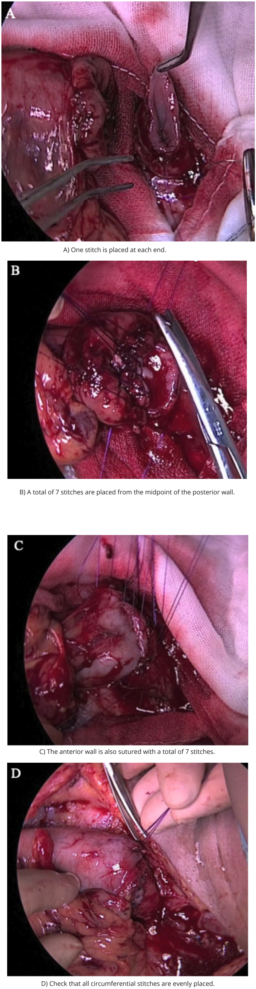
The technique for GA is shown in Figure 1. The anastomosis was performed with 4-0 monofilament absorbable suture. Both anterior and posterior walls were sutured with vertical mattress sutures, and the mucosa was sutured on the lumen side as an introverted anastomosis. The suture should be made with 4-0 absorbable monofilament suture, and 16 stitches per lap should be used for the nodal suture.
One stitch was placed at each end (Fig. 1A). A total of 7 stitches were placed from the midpoint of the posterior wall to ensure even distribution (Fig. 1B). The anterior wall was also sutured with a total of 7 stitches from the center of the anterior wall so that the pitch was even (Fig. 1C). Finally, all circumferential stitches were checked to ensure even placement (Fig. 1D).
2.6 TA method#
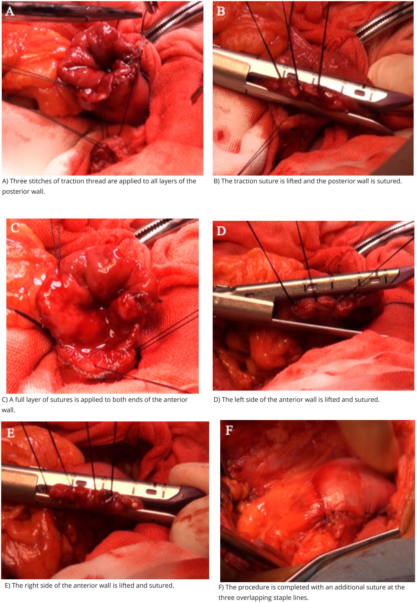
The technique for TA is shown in Figure 2. The anastomosis was performed using three linear staplers. First, three stitches of traction thread were applied to all layers of the posterior wall of the remaining esophagus and gastric tube. To avoid additional entrapment of the anterior wall, one support suture was applied to all layers of the remaining esophagus and gastric conduit (Fig. 2A). The traction suture was lifted, and the posterior wall was sutured with a linear stapler (Fig. 2B). The posterior one-third of the circumference of the posterior wall was sutured as an introverted anastomosis. Next, the left one-third of the anterior wall was sutured. First, a full layer of sutures was applied to both ends of the anterior wall of the esophagus and gastric tube (Fig. 2C). After applying all layers of sutures to the left of the midpoint of two-thirds of the circumference of the anterior wall and the midpoint of the left end of the anterior wall, the three sutures on the left side of the anterior wall were lifted and the left one-third of the circumference of the anterior wall was sutured with a linear stapler (Fig. 2D). This sutured the left one-third of the circumference of the anterior wall as an extroverted anastomosis. Finally, the right one-third of the anterior wall was sutured. The right one-third of the anterior wall was sutured to the external rotation with a linear stapler by placing 2–3 traction sutures in all layers on the right side of the anterior wall and lifting a total of 3–4 traction sutures together with the support suture at the right end of the posterior wall suture line (Fig. 2E). The procedure was completed with an additional suture using 4-0 monofilament absorbable suture at the three overlapping staple lines (Fig. 2F).
2.7 Surgical Outcomes#
The primary endpoint was AL, and secondary endpoints were AS, anastomotic time, and hospital costs. AL was defined as cases with signs of neck wound infection and discharge of purulent effusion or saliva, leakage of contrast from the anastomotic site to the outside of the gastrointestinal tract, and disruption of the anastomotic site on computed tomography, and was assessed using the Clavien–Dindo (CD) classification. AS was assessed by the number of upper gastrointestinal endoscopic dilatations required during the first postoperative year. Anastomotic time was extracted from operating room records. Hospital costs were evaluated based on the difference in the number of points calculated before and after application of the Diagnosis Procedure Combination system.
Perioperative outcomes other than the above endpoints were also examined, such as surgical time, postoperative complications (recurrent nerve palsy and pneumonia), intensive care unit (ICU) stay, and postoperative hospital stay. Postoperative complications were graded according to the CD classification.
2.8 Statistical Analysis#
The χ2 test and Welch’s t-test were used for measures of patient background at surgery and postoperative complications, with both recognizing significant differences at a probability value of 5% or less. Logistic regression analysis, Poisson regression analysis, and linear regression analysis were also performed for primary and secondary endpoints. For logistic regression analyses, odds ratios (ORs) and 95% confidence intervals (CIs) were calculated. For Poisson regression analysis, incident rate ratio and 95%CI were calculated. For linear regression analysis, the coefficient and 95%CI were calculated. All statistical analyses were performed using STATA version 18.0 software (Stata Corporation, College Station, TX, USA).
3 Results#
3.1 Patient Background#
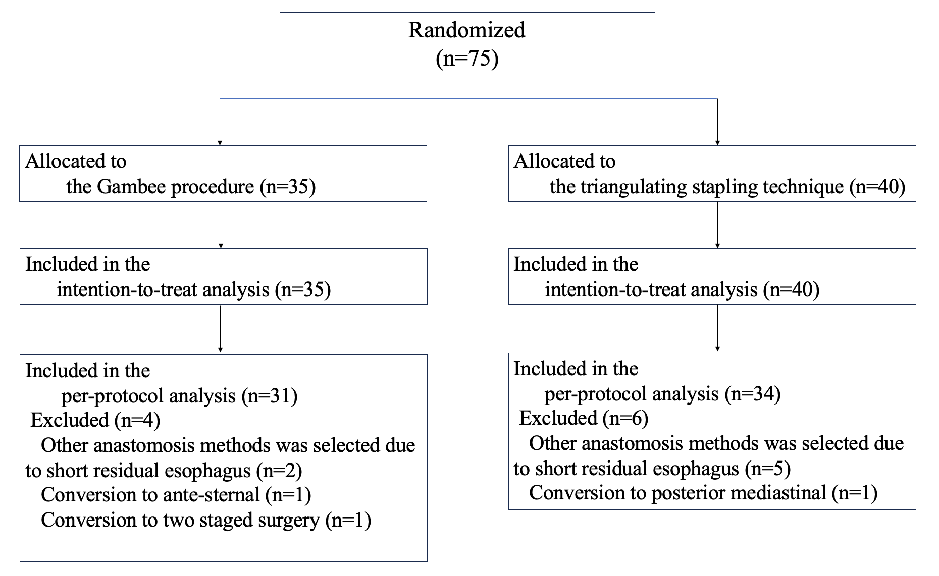
The flow chart of enrolled patients is shown in Figure 3. Seventy-five patients were enrolled from November 2013 to August 2016 and randomized to the GA or TA group. Two patients in the GA group and 5 patients in the TA group underwent a different anastomosis due to shortening of the residual esophagus. One patient in the GA group underwent anterior thoracic reconstruction based on intraoperative findings. One patient in the TA group had a posterior mediastinal route as the reconstruction route based on intraoperative findings, and one patient in the GA group underwent two-stage surgery based on intraoperative findings. These patients were included in the intention-to-treat (ITT) analysis and excluded from the per-protocol-based (PPB) analysis. The ITT population comprised 35 patients in the GA group and 40 patients in the TA group. The PPB population comprised 31 patients in the GA group and 34 patients in the TA group. In both the ITT and PPB populations, the demographic and clinical characteristics of the two groups were similar at baseline (Table 1).
Table 1 Baseline demographic and clinical characteristics of the intention-to-treat population and the per-protocol based population
| Variable | ITT population | PPB population | ||
|---|---|---|---|---|
| the GA group (n=35) | the TA group (n=40) | the GA group (n=31) | the TA group (n=34) | |
| Age (years) | 66.8±9.04 | 65.7±9.15 | 66.6±9.45 | 65.4±9.76 |
| Sex, Male/Female | 26 (74.3)/9 (25.7) | 35 (87.5)/5 (12.5) | 23 (74.2)/9 (25.8) | 29 (85.3)/5 (14.7) |
| BMI (kg/m2) | 21.6 ±3.22 | 22.3±2.51 | 21.6 ±3.19 | 22.1 ±2.67 |
| ASA, 1/2/3/4/5 | 6(17.1)/22(62.9)/6 (17.1)/ 1 (2.9)/0 (0) | 10(25.0)/25(62.5)/ 5 (12.5)/ 0 (0)/0 (0) | 5(16.1)/20(64.5)/5(16.1)/ 1 (3.2)/0 (0) | 10(29.4)/20(58.8)/ 4 (11.8)/ 0 (0)/0 (0) |
| Diabetes mellitus, Yes/No | 7 (20.0)/28 (80.0) | 6 (15.0)/34 (85.0) | 7 (22.6)/24 (77.4) | 3 (8.8)/31 (91.2) |
| Steroid use, Yes/No | 0/(0)/35(100) | 0(0)/40(100) | 0(0)/31(100) | 0(0)/34(100) |
| Histology, SCC/AC/others | 30 (85.7)/4 (11.4)/1 (2.9) | 38 (95.0)/1 (2.5)/1 (2.5) | 26 (83.9)/4 (12.9)/1 (3.2) | 32 (94.1)/1 (2.9)/1 (2.9) |
| Tumor location, Ut/Mt/Lt/Jz | 1(2.9)/17(48.6)/ 13 (37.1)/4 (11.4) | 3(7.5)/21(52.5)/ 15(37.5)/1 (2.5) | 1 (3.2)/15(48.4)/11 (35.5)/ 4 (12.9) | 2(5.9)/19(55.9)/ 12 (35.3)/1 (2.9) |
| P stage category (TNM 8th) 0/ IA/IB/IIA/IIB/IIIA/IIIB/IVA/IVB | 1(2.9)/8(22.9)/5(14.3)/ 0(0)/8(22.9)/1(2.9)/ 4(11.4)/5(14.3)/3(8.6) | 0(0)/6(15.0)/7(17.5)/ 3(7.5)/9(22.5)/3(7.5)/ 7(17.5)/4(10.0)/1(2.5) | 1(3.2)/6(19.4)/3(9.7)/ 0(0)/8(25.8)/1(3.2)/ 4(12.9)/5(16.1)/3(9.7) | 0(0)/4(11.8)/7(20.6)/ 3(8.8)/8(23.5)/2(5.9)/ 6(17.6)/4(11.8)/0(0) |
| Neoadjuvant therapy none (including ESD)/ chemotherapy | 16 (45.7)/19 (54.3) | 17 (42.5)/23 (57.5) | 13 (41.9)/18 (58.1) | 1 5 (44.1)/19 (55.9) |
| Approach to esophagectomy open thoracotomy/ thoracoscopic | 5 (14.3)/30 (85.7) | 4 (10.0)/36 (90.0) | 4 (12.9)/27 (87.1) | 4 (11.8)/30 (88.2) |
| Field lymphadenectomy 2-Field/3-Field | 28 (80.0)/7 (20.0) | 26 (65.0)/14 (35.0) | 25 (80.6)/6 (19.4) | 22 (64.7)/12 (35.3) |
| Laparotomy open laparotomy/mini-laparotomy/HALS | 7 (20.0)/6 (17.1)/22 (62.9) | 5 (12.5)/3 (7.5)/32 (80.0) | 6 (19.4)/6 (19.4)/19 (61.3) | 4 (11.8)/3 (8.8)/27 (79.4) |
| Route of gastric conduit Ante-sternal/Posterior mediastinal/Retro-sternal | 2 (5.7)/0 (0)/33 (94.3) | 0 (0)/1 (2.5)/49 (97.5) | 0 (0)/0 (0)/31(100) | 0 (0)/0 (0)/34(100) |
Values are expressed as mean SD, n (%). AC:adenocarcinoma; ASA:American Society of Anesthesiologist; BMI:body mass index; ESD:endoscopic submucosal dissection; GA: Gambee anastomosis; HALS:hand-assisted laparoscopic surgery; ITT: intention-to-treat; Jz:zone of esophagogastric junction; Lt:lower thoracic; Mt:middle thoracic; PPB: per-protocol based; SCC:squamous cell carcinoma; TA: triangle anastomosis; Ut:upper thoracic.
3.2 Perioperative Outcomes#
Perioperative outcomes were tested by the χ2 test or Welch’s t-test, with the results shown in Table 2. In the ITT population, the incidence of AL was significantly lower in the GA group than in the TA group (p=0.02). Only one patient (2.5%) in the TA group required reoperation for CD grade ≥III. No significant differences were seen in the incidence of AS (GA vs. TA: 31.4% vs. 47.5%, p=0.156). The number of dilatations for AS was significantly lower in the GA group (0.9) than in the TA group (2.7, p=0.0363). Anastomosis time was significantly shorter in the TA group (21.2 min) than in the GA group (25.8 min, p=0.0039). No significant difference was seen in hospital costs (GA vs. TA: 28,272.2 points vs. 31,223.0 points, p=0.4619). Likewise, no significant differences were seen in operative time, recurrent nerve palsy, pneumonia, ICU stay, or postoperative hospital stay (p>0.05). These results were similar in the PPB population, although the incidence of AS was significantly lower in the GA group (29.0%) than in the TA group (50.0%, p=0.005).
Table 2 Perioperative outcomes of the intention-to-treat population and the per-protocol population.
| Variable | ITT population | P value | PPB population | P value | ||
|---|---|---|---|---|---|---|
| The GA group (n=35) | The TA group (n=40) | The GA group (n=31) | The TA group (n=34) | |||
| Primary endpoint | ||||||
| Anastomotic leakage (CD grade) 0 I II III | 30 (85.7) 3 (8.6) 0 (0) 2 (5.7) | 26 (65.0) 1 (2.5) 4 (10.0) 9 (22.5) | 0.02*† | 26 (83.9) 3 (9.7) 0 (0) 2 (6.5) | 21 (61.8) 1 (2.9) 3 (8.8) 9 (26.5) | 0.03*† |
| Secondary endpoints | ||||||
| Anastomotic stenosis Anastomotic stenosis (Number of balloon dilation) Anastomosis time (min) Hospital costs (points) | 11(31.4) 0.9 ±1.49 25.8 ±6.71 28272.2 ±10956.00 | 20 (47.5) 2.7 ±4.90 21.2 ±4.48 31223.0 ±21246.0 | 0.15† 0.03*§ 0.0046**§ 0.44§ | 9 (29.0) 0.8 ±1.41 25.8 ±6.71 28386.3 ±11327.59 | 17 (50.0) 3.0 ±5.21 21.3 ±4.56 30991.6 ±22553.75 | 0.005*† 0.02*§ 0.0051*§ 0.55§ |
| Other outcomes | ||||||
| Surgical time (min) | 593.2 ±81.4 | 613.3 ±62.2 | 0.24§ | 597.1 ±74.2 | 608.7 ±63.68 | 0.50§ |
| Palsy of recurrent laryngeal nerve (CD Grade) 0 I II | 27 (77.1) 6 (17.1) 2 (5.7) | 30 (75.0) 10 (25.0) 0 (0) | 0.24† | 25 (80.6) 5 (16.1) 1 (3.2) | 26 (76.5) 8 (23.5) 0 (0) | 0.46† |
| Pneumonia (CD Grade) 0 I II III | 27 (77.1) 1 (2.9) 7 (20.0) 0 (0) | 29 (72.5) 4 (10.0) 5 (12.5) 2 (5.0) | 0.27† | 25 (80.6) 1 (3.2) 5 (16.1) 0 (0) | 25 (73.5) 4 (11.8) 3 (8.8) 2 (5.9) | 0.244† |
| ICU stay (days) | 6.5 ±1.88 | 6.1 ±2.02 | 0.36§ | 6.5 ±1.81 | 6.3 ±2.11 | 0.65§ |
| Postoperative hospital stay (days) | 22.2 ±12.1 | 26.9 ±16.4 | 0.16§ | 23.0 ±12.63 | 27.8 ±17.11 | 0.21§ |
Values are expressed as mean ±SD, n (%). Chi-square test (χ2 test): †, Welch’s t-test: §, Statistical significance: P-value *<0.05,** <0.005. CD: Clavien-Dindo classification; GA: Gambee anastomosis; ICU: Intensive care unit; ITT: intention-to-treat; PPB: per-protocol based; TA: triangle anastomosis.
3.3 Primary endpoint#
Logistic regression analysis was performed for the incidence of AL (Table 3). In the ITT population, the GA group showed significantly lower frequencies than the TA group for CD grade ≥I and CD grade ≥II, but not for CD grade ≥III (CD ≥I: OR 3.231, 95%CI 1.025–10.186, p=0.045; CD ≥II: OR 7.944, 95%CI 1.648–38.308, p=0.01; CD ≥III: OR 4.790, 95%CI 0.95878–23.934, p=0.056). In the PPB population, no significant difference was seen for CD grade ≥I, but the GA group showed a significantly lower rate than the TA group for CD grade ≥II and CD grade ≥III (CD ≥I: OR 3.22, 95%CI 0.988–10.485, p=0.052; CD ≥II: OR 7.91, 95%CI 1.603–39.026, p=0.011; CD ≥III: OR 5.22, 95%CI 1.030–26.453, p=0.046).
Table 3 Primary endpoint.
| CD grade of anastomotic leakage | The GA group | The TA group | Odds Ratio | 95%CI | P value |
|---|---|---|---|---|---|
| ITT population | n =35 | n=40 | |||
| CD≥I None | 30 (85.7) | 26 (65.0) | |||
| CD≥I | 5 (14.3) | 14 (35.0) | 3.231 | 1.025- 10.186 | 0.04 |
| CD≥II None | 33 (94.3) | 27 (67.5) | |||
| CD≥II | 2 (5.7) | 13 (32.5) | 7.944 | 1.648- 38.308 | 0.01* |
| CD≥III None | 33 (94.3) | 31(77.5) | |||
| CD≥III | 2 (5.7) | 9 (22.5) | 4.790 | 0.95878- 23.934 | 0.05 |
| PPB population | n=31 | n=34 | |||
| CD≥I None | 26 (83.9) | 21 (61.8) | |||
| CD≥I | 5 (16.1) | 13 (38.2) | 3.22 | 0.988 - 10.485 | 0.05 |
| CD≥II None | 29 (93.5) | 22 (64.7) | |||
| CD≥II | 2 (6.5) | 12 (35.3) | 7.91 | 1.603- 39.026 | 0.01* |
| CD≥III None | 29 (93.5) | 25 (73.5) | |||
| CD≥III | 2 (6.5) | 9 (26.5) | 5.22 | 1.030 - 26.453 | 0.04* |
Values are expressed as mean ±SD, n (%). Logistic regression. Statistical significance: P-value *<0.05. CD: Clavien-Dindo classification; GA: Gambee anastomosis; ITT: intention-to-treat; PPB: per-protocol based; TA: triangle anastomosis.
3.4 Secondary endpoints#
Poisson regression analysis was performed for the number of dilatations for AS and linear regression analysis for anastomotic time and hospital costs (Table 4). In the ITT population, the number of dilatations for AS was significantly lower in the GA group than in the TA group (incident rate ratio 3.077, 95%CI 2.064–4.585, p<0.001). Anastomosis time was significantly shorter in the TA group than in the GA group (coefficient -4.573, 95%CI -7.609– -1.537, p=0.004). No significant differences were apparent in hospital costs (coefficient 2950.7, 95%CI -4899.362–10,901.26, p=0.462). Results were similar for the PPB population.
Table 4 Secondary endpoint.
| the GA group | the TA group | IRR/Coefficient | 95%CI | P value | |
|---|---|---|---|---|---|
| ITT population | n=35 | n=40 | |||
| Anastomotic stenosis | |||||
| (Number of balloon dilation) | 0.9 ±1.49 | 2.7 ±4.90 | 3.077a | 2.064 - 4.585 | <0.001** |
| Anastomosis time (min) | 25.8 ±6.71 | 21.2 ±4.48 | -4.573b | -7.609 - -1.537 | 0.004** |
| Hospital costs (points) | 28272.2 ±10956.00 | 31223.0 ±21246.0 | 2950.7b | -4899.362 - 10901.26 | 0.46 |
| PPB population | n=31 | n=34 | |||
| Anastomotic stenosis | |||||
| (Number of balloon dilation) | 0.8 ±1.41 | 3.0 ±5.21 | 3.913a | 2.509 - 6.102 | <0.001** |
| Anastomosis time (min) | 25.8 ±6.71 | 21.3 ±4.56 | -4.564b | -7.657 - -1.473 | 0.005* |
| Hospital costs (points) | 28386.3 ±11327.59 | 30991.6±22553.75 | 2605.2b | -6376.163 - 11586.64 | 0.56 |
Values are expressed as mean ±SD, n (%). Anastomotic stenosis; Poisson regression, Anastomosis time and Hospital costs; Linear regression, a; IRR, b: Coefficient, Statistical significance: P-value *<0.05, **<0.005. CI: confidence intervals; GA: Gambee anastomosis; IRR: Incident Rate Ratio; ITT: intention-to-treat; PPB: per-protocol based; TA: triangle anastomosis.
4 Comment#
The present RCT compared two methods of cervical esophagogastric tube anastomosis after subtotal esophagectomy. The GA group showed a significantly lower incidence of AL (CD grade ≥II) and the number of postoperative dilatations for AS within one year compared to the TA group. On the other hand, anastomosis time was significantly shorter in the TA group than in the GA group. No significant differences in hospital costs or other perioperative outcomes were evident between groups.
Furukawa et al. reported a retrospective study comparing TA, circular stapler and hand-sewn anastomoses11 . Toh et al. reported a retrospective study comparing TA and hand-sewn anastomoses14 . Both reported that TA had a lower incidence of AL and AS than hand-sewn anastomosis.
Regarding AL, the incidence of CD grade ≥II was significantly lower in the GA group in this study, at 5.7% (ITT) in the GA group and 32.5% (ITT) in the TA group. Previous reports have shown AL rates of 1.1–27.3% for hand-sewn procedures and 2–17.9% for TA. Although direct comparison of our results with reports from other institutions is difficult due to inconsistent definitions, we should conclude that the rate of suture failure is high in the TA group. Factors contributing to AL include comorbidities, organ perfusion and tension, and surgeon skill. Regarding comorbidities, both groups were assumed to be equally assigned by stratified randomization. Regarding the method of evaluation of organ blood flow, differences were considered unlikely to occur because the surgeon was limited to one person. Although the same surgeon was careful not to increase the tension applied to the anastomosis site, the TA method involves three stitches of traction thread followed by suturing with a linear stapler, so tension may be higher than assumed at the start of the anastomosis. Regarding surgeon skill, both GA and TA in this study were performed by a surgeon with sufficient experience in both methods. The advantage of mechanical anastomosis is that the technique is easy to standardize and less prone to technical errors19 , but as with hand-sewn methods, knowledge and skill are required regarding tips and pitfalls for each technique. In this study, we performed TA in which the posterior wall was sutured with an introverted suture and the remaining two sides were sutured with an extroverted suture. A risk of AL is present at the overlap of the in- and out-staples with this method. The double-stapled area was buried with an additional suture, and this buried suture may have increased the tension applied to the area. More experience and knowledge of the details, including the consideration of the tension applied to the anastomosis, are thus necessary.
Regarding AS, the number of procedures was significantly lower in the GA group in this study. The percentage of patients requiring dilation was 31.4% (ITT) in the GA group and 47.5% (ITT) in the TA group. Previous studies have reported AS rates of 2.3–58% for hand-sewn and 8.3–27.5% for TA. Although direct comparison of the results from this study with those of previous reports is difficult given the lack of uniformity in definitions, we should conclude that the AS rate was relatively high in the TA group. AL has been reported as a risk factor for AS27 , and the AL rate was higher in the TA group in this study, which may have resulted in more AS.
The advantages of mechanical anastomosis include a reduced surgical time10,11,15,16,18,20 . Few studies have compared anastomosis times, but Furukawa et al. reported mean times of 54 ± 18 min for a hand-sewn technique and a shorter 24.0 ± 11 min for TA. In the present study, the anastomosis time was significantly shorter in the TA group. However, the mean difference in anastomosis time in this study was approximately 4 min, and no significant difference in surgical time was evident between groups, making the significant difference in anastomosis time of little clinical significance.
Cost is an issue for mechanical anastomosis13,21,26 , but we could not find any reports comparing anastomosis methods from a health economics point of view. In the present study, we compared the costs of hospitalization and found no significant difference between groups. This may be because although the AL rate was higher in the TA group, almost all patients recovered with conservative treatment, and postoperative hospital stay thus did not differ significantly.
Based on the results of this study, we have adopted GA for end-to-end anastomotic reconstruction after subtotal esophagectomy. However, we believe that the anastomosis method most familiar to each institution and surgeon is most appropriate.
This RCT showed several limitations. First, data were collected from a small cohort at a single center. Second, we were unable to examine outpatient procedures and readmissions from a health economic perspective. To address these issues, we believe that a multicenter prospective RCT with long-term outcomes should be performed.
In conclusion, we believe that the GA is superior to the TA in terms of AL and AS for esophagogastric neck anastomosis after subtotal esophagectomy.
Declarations
€Funding
◎Competing interests
♥Acknowledgements
Article information
- Published
Copyright © 2025 Dr. Kazuhiro Noma, M.D. Ph.D.. This is an open access article distributed under the Creative Commons Attribution License, which permits unrestricted use, distribution, and reproduction in any medium, provided the original work is properly cited. CC BY 4.0 · Corrections policy
References 27#
- 1
Chen MF, Yang YH, Lai CH, Chen PC, Chen WC. Outcome of patients with esophageal cancer: a nationwide analysis. Ann Surg Oncol. 2013;20(9):3023-3030. doi:10.1245/s10434-013-2935-4
- 2
Akutsu Y, Kato K, Igaki H, et al. The Prevalence of Overall and Initial Lymph Node Metastases in Clinical T1N0 Thoracic Esophageal Cancer: From the Results of JCOG0502, a Prospective Multicenter Study. Ann Surg. 2016;264(6):1009-1015. doi:10.1097/SLA.0000000000001557
- 3
Law S, Fok M, Chu KM, Wong J. Comparison of hand-sewn and stapled esophagogastric anastomosis after esophageal resection for cancer: a prospective randomized controlled trial. Ann Surg. 1997;226(2):169-173. doi:10.1097/00000658-199708000-00008
- 4
Beitler AL, Urschel JD. Comparison of stapled and hand-sewn esophagogastric anastomoses. Am J Surg. 1998;175(4):337-340. doi:10.1016/s0002-9610(98)00002-6
- 5
Orringer MB, Marshall B, Iannettoni MD. Eliminating the cervical esophagogastric anastomotic leak with a side-to-side stapled anastomosis. J Thorac Cardiovasc Surg. 2000;119(2):277-288. doi:10.1016/S0022-5223(00)70183-8
- 6
Singh D, Maley RH, Santucci T, et al. Experience and technique of stapled mechanical cervical esophagogastric anastomosis. Ann Thorac Surg. 2001;71(2):419-424. doi:10.1016/s0003-4975(00)02337-7
- 7
Urschel JD, Blewett CJ, Bennett WF, Miller JD, Young JE. Handsewn or stapled esophagogastric anastomoses after esophagectomy for cancer: meta-analysis of randomized controlled trials. Dis Esophagus. 2001;14(3-4):212-217. doi:10.1046/j.1442-2050.2001.00187.x
- 8
Alanezi K, Urschel JD. Mortality secondary to esophageal anastomotic leak. Ann Thorac Cardiovasc Surg. 2004;10(2):71-75.
- 9
Ercan S, Rice TW, Murthy SC, Rybicki LA, Blackstone EH. Does esophagogastric anastomotic technique influence the outcome of patients with esophageal cancer?. J Thorac Cardiovasc Surg. 2005;129(3):623-631. doi:10.1016/j.jtcvs.2004.08.024
- 10
Hsu HH, Chen JS, Huang PM, Lee JM, Lee YC. Comparison of manual and mechanical cervical esophagogastric anastomosis after esophageal resection for squamous cell carcinoma: a prospective randomized controlled trial. Eur J Cardiothorac Surg. 2004;25(6):1097-1101. doi:10.1016/j.ejcts.2004.02.026
- 11
Furukawa Y, Hanyu N, Hirai K, et al. Usefulness of automatic triangular anastomosis for esophageal cancer surgery using a linear stapler (TA-30). Ann Thorac Cardiovasc Surg. 2005;11(2):80-86.
- 12
Blackmon SH, Correa AM, Wynn B, et al. Propensity-matched analysis of three techniques for intrathoracic esophagogastric anastomosis. Ann Thorac Surg. 2007;83(5):1805-1813. doi:10.1016/j.athoracsur.2007.01.046
- 13
Zhu ZJ, Zhao YF, Chen LQ, et al. Clinical application of layered anastomosis during esophagogastrostomy. World J Surg. 2008;32(4):583-588. doi:10.1007/s00268-007-9396-5
- 14
Toh Y, Sakaguchi Y, Ikeda O, et al. The triangulating stapling technique for cervical esophagogastric anastomosis after esophagectomy. Surg Today. 2009;39(3):201-206. doi:10.1007/s00595-008-3827-2
- 15
Kim RH, Takabe K. Methods of esophagogastric anastomoses following esophagectomy for cancer: A systematic review. J Surg Oncol. 2010;101(6):527-533. doi:10.1002/jso.21510
- 16
Honda M, Kuriyama A, Noma H, Nunobe S, Furukawa TA. Hand-sewn versus mechanical esophagogastric anastomosis after esophagectomy: a systematic review and meta-analysis. Ann Surg. 2013;257(2):238-248. doi:10.1097/SLA.0b013e31826d4723
- 17
Markar SR, Arya S, Karthikesalingam A, Hanna GB. Technical factors that affect anastomotic integrity following esophagectomy: systematic review and meta-analysis. Ann Surg Oncol. 2013;20(13):4274-4281. doi:10.1245/s10434-013-3189-x
- 18
Castro PM, Ribeiro FP, Rocha Ade F, Mazzurana M, Alvarez GA. Hand-sewn versus stapler esophagogastric anastomosis after esophageal ressection: systematic review and meta-analysis. Arq Bras Cir Dig. 2014;27(3):216-221. doi:10.1590/s0102-67202014000300014
- 19
Deng XF, Liu QX, Zhou D, Min JX, Dai JG. Hand-sewn vs linearly stapled esophagogastric anastomosis for esophageal cancer: a meta-analysis. World J Gastroenterol. 2015;21(15):4757-4764. doi:10.3748/wjg.v21.i15.4757
- 20
Liu QX, Qiu Y, Deng XF, Min JX, Dai JG. Comparison of outcomes following end-to-end hand-sewn and mechanical oesophagogastric anastomosis after oesophagectomy for carcinoma: a prospective randomized controlled trial. Eur J Cardiothorac Surg. 2015;47(3):e118-e123. doi:10.1093/ejcts/ezu457
- 21
Wang Q, He XR, Shi CH, et al. Hand-Sewn Versus Stapled Esophagogastric Anastomosis in the Neck: A Systematic Review and Meta-analysis of Randomized Controlled Trials. Indian J Surg. 2015;77(2):133-140. doi:10.1007/s12262-013-0984-3
- 22
Mishra PK, Shah H, Gupta N, et al. Stapled versus hand-sewn cervical esophagogastric anastomosis in patients undergoing esophagectomy: A Retrospective Cohort Study. Ann Med Surg (Lond). 2016;5:118-124. Published 2016 Jan 4. doi:10.1016/j.amsu.2015.12.063
- 23
Kamarajah SK, Bundred JR, Singh P, Pasquali S, Griffiths EA. Anastomotic techniques for oesophagectomy for malignancy: systematic review and network meta-analysis. BJS Open. 2020;4(4):563-576. doi:10.1002/bjs5.50298
- 24
Nederlof N, Tilanus HW, de Vringer T, et al. A single blinded randomized controlled trial comparing semi-mechanical with hand-sewn cervical anastomosis after esophagectomy for cancer (SHARE-study). J Surg Oncol. 2020;122(8):1616-1623. doi:10.1002/jso.26209
- 25
Chen B, Xia P, Tang W, Huang S. Which Anastomotic Techniques Is the Best Choice for Cervical Esophagogastric Anastomosis in Esophagectomy? A Bayesian Network Meta-Analysis. J Gastrointest Surg. 2023;27(2):422-432. doi:10.1007/s11605-022-05482-y
- 26
Ozawa H, Kawakubo H, Nishimura E, et al. Comparison of hand-sewn and circular stapled esophagogastric anastomoses in the neck after esophagectomy for thoracic esophageal cancer: a propensity score-matched analysis. Dis Esophagus. 2023;36(3):doac066. doi:10.1093/dote/doac066
- 27
Zhong Y, Sun R, Li W, et al. Risk factors for esophageal anastomotic stricture after esophagectomy: a meta-analysis. BMC Cancer. 2024;24(1):872. Published 2024 Jul 19. doi:10.1186/s12885-024-12625-8
Cited by 1#
- The Use of Barbed Sutures in Free Jejunal TransferS. Higuchi, M. Kambe, K. Ebisawa et al.Journal of Plastic and Reconstructive Surgery · 2026 · doi.org/10.53045/jprs.2025-0148
Each citation is confirmed in the citing article's own reference list (Crossref).