Successful Management Of Cheilitis Glandularis With Intralesional Corticosteroid Injection: A Case Report
Norberto Sugaya DDS
Norberto Sugaya DDS- Department of Stomatology, Division of Oral Medicine, School of Dentistry, University of Sao Paulo, Brazil.
Dante Migliari DDS
Dante Migliari DDS- Department of Stomatology, Division of Oral Medicine, School of Dentistry, University of Sao Paulo, Brazil.
Show affiliations (1)
- Department of Stomatology, Division of Oral Medicine, School of Dentistry, University of Sao Paulo, Brazil.
Published
Abstract
Cheilitis glandularis (CG) is an inflammatory condition of unknown cause, with its etiopathogenic events involving predominantly the minor salivary glands of the lips. Early characterization of the disease is based on the presence of a recurrent, nonadherent crust-like plaque formation mainly on the lower lip mucosal, a finding considered a hallmark sign of the disease. While CG is relatively straightforward to diagnose, its treatment is a challenge. The article highlights the clinical presentation of the disease and details a case in which successful management could only be achieved with intralesional injection of corticosteroids after initial attempts using moderate to potent corticosteroids or immunosuppressor topical ointments failed to provide sustained benefit.
1 Introduction#
Cheilitis glandularis (CG) is a rare inflammatory condition of unknown cause that primarily affects the minor salivary glands of the lips while also involving the surrounding tissues as the inflammation progresses. The disease predominantly affects the lower lip rather than the upper one. CG occurs more often in adults (> 40 years old) than in young and affects almost exclusively white individuals. It primarily affects males, with male/female ratio of 3:1. The CG seems to occur at random—no specific triggering factor preceding its onset has been identified to date[1,2].
The clinical features of CG are reasonably specific to the disease, making its diagnosis relatively straightforward. In terms of clinical etiopathogenic basis, the labial mucosa exhibits a dilation of the orifices of the minor salivary glands, through which a thick saliva (mucin-rich) flows more intensely than normal due to the inflammation of the glandular parenchyma. This excessive salivary flow eventually abates, leading to the development of yellowish plaques (or crusts) that spread over the external area of labial mucosa surface and part of the vermilion border. These plaques are easily removed, but they repeatedly re-form, mostly when the patient is asleep. Along with these plaques, a patient commonly develops an enlargement and eversion of the lip(s)[1-3].
Managing CG remains a challenge; there are no guidelines to deal with this condition efficiently. This article reports a case of CG successfully managed by intralesional steroid suspension injection following unsuccessful attempts to treat this condition by applying either topical steroids or immunosuppressor ointments.
2 Case Report#
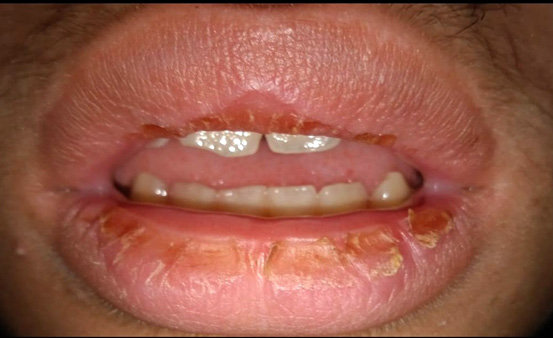
A 15-year-old white male was referred to our oral medicine clinic for a diagnosis and treatment of recurrent yellowish crusts of his both lips that had been present for four months. The patient reported that he normally noted these crusts in the morning when he woke up (Fig.1). These crusted lesions were not adherent and were easily removed with warm water but reappeared almost every morning. He also presented an enlargement of both lips and eversion of the lower lip. The esthetic and functional effects of these lesions were affecting the patient’s well-being, but he was otherwise a healthy person.
3 Differential Diagnosis#
The diagnosis of the present case was made exclusively on a clinical basis after other diseases that may exhibit clinical features similar to those of CG, such as contact cheilitis and cheilitis granulomatosa, were ruled out. Specifically, contact cheilitis manifests by adherent, non-removable scales rather than crusted lesions, and appears more prominently on the lip vermilion, while an allergic contactant can usually be identified[4]. Cheilitis granulomatosa, in turn, despite sharing some similarities with CG concerning lip swelling and eversion[5], also lacks the crust formation on the lips, which is almost the hallmark for a diagnosis of CG. Additionally, biopsy has not been a standard method to determine the diagnosis in most CG cases, since its histopathological findings, according to two relevant studies on this issue, are nonspecific, consisting mainly of chronic inflammation with various degrees of sialadenitis, along with ductal ectasia of the minor salivary glands and fibrosis within the glands. Therefore, a biopsy wouldn’t be helpful for this patient in terms of diagnosis and further management.
4 Management#
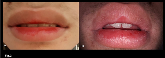
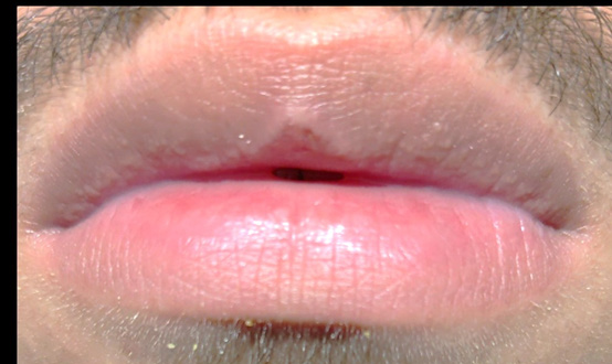
Based on a few studies and our own knowledge in dealing with this type of lip manifestations[2,3,6], the patient was initially treated with medium-potency topical corticosteroids in combination with an antibiotic (betamethasone dipropionate 0.5 mg/g + gentamicin sulfate 1.0 mg/g), applied twice a day for approximately 2–4 weeks). This association appears to enhance the benefits of steroids by reducing bacterial colonization on the lesion’s surface through the effects of the antibiotic. With this treatment, some improvement in the patient’s condition was observed— the plaques no longer reappeared daily. After three weeks, the medication was tapered and withdrawn. Unfortunately, recurrence occurred 1 week later. The patient was then switched first to tacrolimus ointment (0.1%) and later to clobetasol propionate ointment (0.05%). However, both of these medications induced skin and mucosa reactions (redness and burning sensation, Fig. 2A and B) and were withdrawn. Another attempted to treat the condition involved a single intralesional steroid injection into each lip. Specifically, a 5mg/mL betamethasone dipropionate suspension was used on both lips. Prior to the steroid injection, the patient was given local anesthesia using a 2% lidocaine with epinephrine to reduce pain during the intralesional injection and to facilitate accurate placement of the medication inside the lip tissue. This treatment approach resulted in substantial reduction of the severity of the lip lesions by eliminating the crust formation and reestablishing the lip anatomy. The benefits of a single steroid injection in each lip were long-lasting, with no recurrence developing over a four-year follow-up (Fig 3).
5 Discussion#
CG is an uncommon lip condition in a clinical setting. When cases emerge, some difficulty in achieving treatment success can occur given the refractoriness of CG to the current standard therapies. Accordingly, the esthetic and functional impairments associated with CG require novel treatment options[3,6,7].
At present, CG is mainly managed using topical medication. Systemic medication, mostly corticosteroids, are also available and might produce some improvement, but their long-term use is not without a risk. In fact, their side effects, mainly hypertension, higher risk of infections, insomnia, and hyperglycemia, usually outweigh their possible benefit. Meanwhile, when medications such as moderate to potent corticosteroids and immunosuppressors such as tacrolimus are applied topically, they may achieve some improvements but usually fail to permanently resolve the symptomatology, while relapsing tends to occur once the medication is withdrawn. Topical agents (potent corticosteroids or immunosuppressors) can also cause skin and mucosal reactions before any benefit is seen[7].
In the present case, long-term benefits were achieved only with the intralesional injection of corticosteroids. Other treatment options attempted, such as the use of ointments containing moderate to potent corticosteroids and immunosuppressor, failed to effectively control the disease’s clinical manifestation.
6 Conclusion#
This study suggests that intralesional injection of corticosteroids is an appropriate therapeutic option in refractory cases of CG.
Article information
- Published
Copyright © 2026 Dante Migliari. This is an open access article distributed under the Creative Commons Attribution License, which permits unrestricted use, distribution, and reproduction in any medium, provided the original work is properly cited. CC BY 4.0 · Corrections policy
References 7#
- 1
Reiter S, Vered M, Yarom N, et al., “Cheilitis glandularis: clinico-histopathological diagnostic criteria,” Oral Diseases 3 (2011): 335-9. doi: 10.1111/j.1601-0825.2010.01762.
- 2
Nico MM, Nakano de Melo J, Lourenço SV, “Cheilitis glandularis: a clinicopathological study in 22 patients,” J Am Acad Dermatol 62 (2010):233-8. doi: 10.1016/j.jaad.2009.06.038.
- 3
Sugaya N, Migliari D, “Cheilitis Glandularis of Both Lips: Successful Treatment with a Combination of an Intralesional Steroid Injection and Tacrolimus Ointment,” Case Reports in Dentistry 18 (2018):9169208. doi: 10.1155/2018/9169208.
- 4
Han J, Warshaw EM, “Allergic Contact Cheilitis due to Candelilla Wax,” Dermatitis 34 (2023) :548-550. doi: 10.1089/derm.2023.0062.
- 5
van der Waal RI, Schulten EA, van der Meij EH, van de Scheur MR, Starink TM, van der Waal I, “Cheilitis granulomatosa: overview of 13 patients with long-term follow-up--results of management,” International Journal of Dermatology 41 (2002) :225-9. doi: 10.1046/j.1365-4362.2002.01466.x.
- 6
Bovenschen HJ, “Novel treatment for cheilitis glandularis,” Acta Dermato-Venereologica 89 (2009): 99-100. doi: 10.2340/00015555-0557.
- 7
Erkek E, Sahin S, Kilic R, et al., “A case of cheilitis glandularis superimposed on oral lichen planus: successful palliative treatment with topical tacrolimus and pimecrolimus,” Journal of the European Academy of Dermatology and Venereology 21 (2007): 999-1000. doi: 10.1111/j.1468-3083.2007.02071.x.