Full Text Hide / show
Introduction
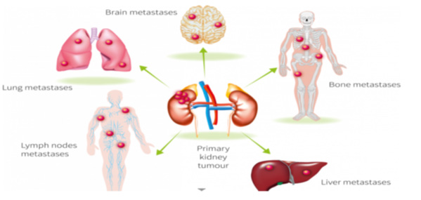
Worldwide, renal cell carcinoma (RCC) represents the sixth most frequently diagnosed cancer in men and the 10th in women, accounting for 5% and 3% of all oncological diagnoses, respectively (1). There are several histological subtypes including clear cell renal cell carcinoma (ccRCC) and papillary renal cell carcinoma (pRCC) (2). The natural history of RCC depends on a variety of factors, and metastatic rates increase with tumour size; in ccRCC, metastatic rates for tumours ≤4 cm, 4–≤7 cm, 7–≤10 cm, and >10 cm were 2.1%, 8.8%, 24.6%, and 41.9%, respectively, compared with 1.7%, 6.7%, 16.0%, and 28.6% for pRCC(3). The most common sites of metastasis in RCC are the lungs (45.2%), bone (29.5%), lymph nodes (21.8%), liver (20.3%), adrenal glands (8.9%) and brain (8.1%) (4).
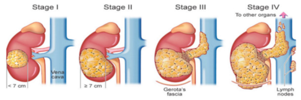
Image 1 below displays the stages of renal cancer and degree of invasion (5), and Image 2 shows the various routes and sites of metastatic spread in RCC (6). Abstract The management of small renal masses (SRM) has been evolving over recent years. There is still debate over whether active surveillance can replace radical nephrectomy. We present the case of a woman in her early 90s with an 18cm renal mass that developed over 18 years. The patient was referred to the medical assessment unit. Clinical examination revealed a palpable right upper quadrant mass. A subsequent CT scan of the abdomen and pelvis confirmed a 13cmx17cmx18cm mass arising from the right kidney.
The woman had a past medical history of colon adenocarcinoma 18 years prior to this presentation. A CT scan performed post-surgery that year showed an incidental 1.7cm right kidney lesion. This woman was discussed at the Urology MDM. The renal lesion was classified as a Bosniak IV renal mass, indicating it was radiologically malignant. The patient did not wish to undergo any intervention. When last reviewed virtually this year, the patient was well, independent and asymptomatic. A SRM is defined as an incidentally detected contrast enhancing or cystic lesion that is ≤4cm. Among surgically treated SRMs, 80% are malignant; although most are low-grade tumours, the remaining 20% are benign. According to American Society of Clinical Oncology Guidelines, active surveillance is reasonable for smaller renal lesions in older patients, those with multiple comorbidities, or those who have a limited life expectancy.
This patient displayed the natural history of a renal cell carcinoma over an 18-year period. Despite the tumour growth, the patient remained asymptomatic with preserved renal function. Image 1. The stages of RCC and degree of invasion (5). Overall five-year survival rates for stage I is 90%; stage II and III is 75% and stage IV is 15%. Image 2. The routes and sites of metastases in RCC (6). The incidence of renal malignancies has increased in recent years due to the increase in incidental findings of small renal masses. The preferred treatment is surgical in young, fit patients, however in older patients with notable co-morbidities there is a role for ablation or active surveillance.
In this case report, we examine the natural history of a small renal mass in a patient that did not undergo any interventions. See Table 1 below outlining some key definitions relevant to this case. Table 1. Key radiological definitions. Definitions Small renal mass (SRM) An incidentally detected contrast enhancing or cystic lesion that is 4cm. Bosniak IV lesion Complex cystic renal mass with a solid enhancing component indicating a high probability of malignancy.
Case Presentation
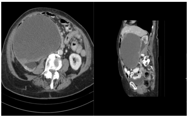
A woman in her 90s was referred to the local medical assessment unit (MAU) with resolved right sided motor weakness of her hand and leg. She was otherwise well with no systemic symptoms, including weight loss, fatigue or night sweats. Clinical examination revealed no focal neurology but there was a palpable right upper quadrant mass. The patient underwent an abdominal ultrasound which reported a large vascular mass inferior to the liver. It measured 19.7cm x 12.9cm. A subsequent CT scan of the abdomen and pelvis confirmed a 13cmx17cmx18cm mass arising from the anterior aspect of the right kidney. The patient was diagnosed with colon adenocarcinoma 18 years ago.
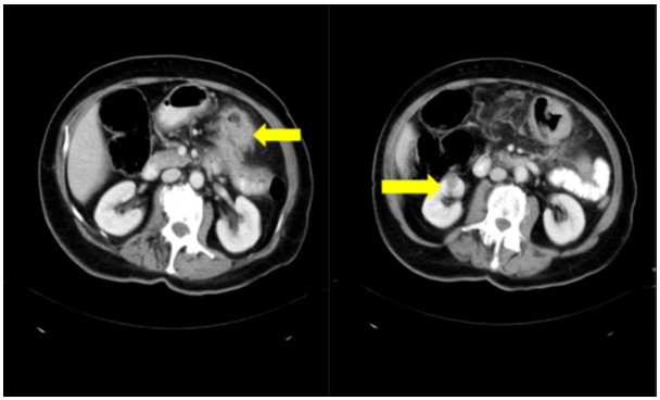
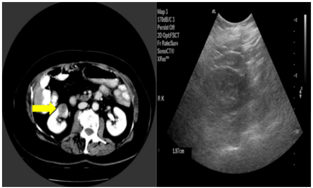
Her staging CT scan showed a transverse colon lesion and a right renal lesion (that was not initially reported), and she underwent a hemi-colectomy followed by adjuvant chemotherapy. A re-staging CT scan performed post-surgery and pre-chemotherapy later that year reported the incidental 1.7cm right kidney lesion and no evidence of metastatic disease. The kidney was further evaluated by ultrasound which showed a 1.9cm cystic renal lesion on the lower pole of the right kidney. Image 3. CT scan showing colon adenocarcinoma (left) and unreported right renal lesion (right). Image 4. Re-staging CT scan post-operatively and pre-chemotherapy showing renal cyst 1.7cm (left) and renal ultrasound showing 1.9cm renal lesion (right) two months following Image 1.
The rest of this patient’s past medical history includes uterine fibroids, hypertension, atrial fibrillation and gout. She has one sister and two brothers, one of whom had renal cell carcinoma in the past. She lives alone independently and is a non-smoker with minimal alcohol intake. The patient underwent numerous abdominal and pelvic CT scans over a four-year period as per surveillance of colon cancer. The last CT 14 years before presenting to the MAU showed that the right kidney lesion measured 4cm and appeared more solid than cystic. She underwent two subsequent renal ultrasounds, one 13 years before, confirming the 4cm solid lesion from lower pole of right kidney, and another 12 years before which showed a stable 3.9cm lesion in lower pole of right kidney.
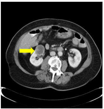
Image 5. Surveillance CT scan showing stable 4cm renal lesion 4 years following Image 1. The patient underwent no further imaging until she was referred to the MAU for investigation of her neurological symptoms 18 years later. Her CT brain was normal; however, the CT abdomen and pelvis showed that the right renal lesion was now 18cm, predominantly cystic with solid components. There was no invasion of surrounding structures or pathologically enlarged lymph nodes, although mass effect was noted. Her renal function was normal with an eGFR >90 ml/min/1.73m2. Her chest radiograph was normal. Image 6. Axial CT scan (left) and sagittal CT scan (right) showing 18cm renal lesion 18 years following Image 1.
This woman was discussed at the Urology MDM. The renal lesion was radiologically classified as a Bosniak IV renal mass, indicating it was highly likely to be malignant. The results were discussed with the patient and her family. The patient did not wish to undergo any intervention, including surgery. A conservative approach has been taken with no further intervention. She last reviewed virtually this year (two years since Image 4), and she was well, independent and asymptomatic.
Discussion
A SRM is defined as an incidentally detected contrast enhancing or cystic lesion that is ≤4 cm (7). Among surgically treated SRMs, 80% are malignant; although most are low-grade tumours, the remaining 20% are benign (8). SRMs are best evaluated with CT or MRI. They are characterized as predominantly cystic or solid. The Bosniak Classification system, as outlined in Table 2, is then used to stratify the risk of malignancy in cystic masses from I to IV. A Bosniak IV lesion has a 90% chance of being malignant (9). Table 2. Description of Bosniak Classification Bosniak Classification CT Features Risk of Malignancy Recommended Management I Simple cyst with thin wall No septae, calcifications or enhancement Water attenuation (-10 to 20 HU) None No therapy or follow-up II Few hair-line septae Fine calcifications No enhancement May be high attenuation (>20HU) Cyst <3cm 0-6% No therapy or follow-up IIF Multiple hairline septae Minimally smooth thickening of septae or wall Can have calcifications Intrarenal attenuation >20HU Cyst >3cm No enhancement 50% Surveillance IV Enhancing soft tissue components independent of wall or septum 75-90% Partial or radical nephrectomy Active surveillance and nephron-sparing approaches are now accepted options for primary treatment modalities for small renal masses (≤4 cm) (10).
Our patient initially had a 1.7cm renal lesion which underwent surveillance for six years. It grew to exactly 4cm after which she was lost to follow-up. According to American Society of Clinical Oncology (ASCO) Guidelines, active surveillance is reasonable for smaller renal lesions in older patients, those with multiple or major co-morbidities, high surgical risk, or those who have a limited life expectancy (11). The National Comprehensive Cancer Network (NCCN) currently recommends partial nephrectomy, but with consideration for active surveillance or ablation for stage I RCC (12). Another study found there was only one cancer specific death in 332 patients with a complex renal cyst, with no cancer deaths among the patients who did not undergo any intervention (13).
Table 3 gives a brief overview of current recommendations for the management of SRMs from ASCO and the NCCN. Table 3. Summary of current guidelines on the management of SRMs. ASCO Recommendations NCCN Recommendations Biopsy All patients with a SRM should undergo biopsy unless contraindications exist Consider if for surgery or active surveillance, needed prior to ablation Active surveillance For patients with significant co-morbidities, limited life expectancy (<5 years), SRM ≤1cm For masses <3cm, T1a tumours ≤4 cm and in patients with co-morbidities Surgical management Partial nephrectomy preferred to radical nephrectomy by both guidelines to preserve renal function Partial nephrectomy as the standard of treatment for SRMs in surgical fit patients For renal cell carcinoma stage II-IV, with the option of stereotactic body radiation therapy instead for stage II and III Ablative management Should be considered for patients in whom complete ablation can be achieved For older patients, co-morbid patients or T1b masses not eligible for surgery In our patient these guidelines would have supported conservative management at the time of initial discovery of her renal lesion.
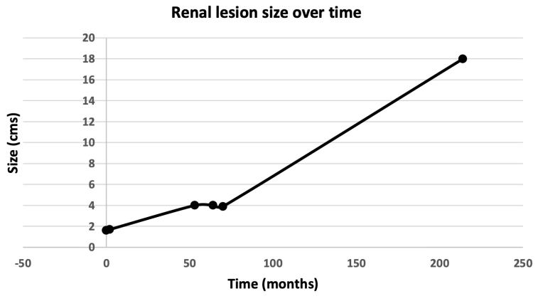
Although her renal mass grew from 1.7cm to 18cm over 18 years, there was no invasion of surrounding structures, lymph node involvement or evidence of metastatic spread. The ‘growth’ of the renal lesion is displayed on Figure 1 below. The patient also remains asymptomatic with normal renal function. Despite the remarkable size of her Bosniak IV renal lesion, the patient has not come to any harm. Figure 1. The growth of the renal lesion over time. However, there are risks of having a large renal cystic mass, such as mass effect causing abdominal discomfort, possibly obstruction and a rare complication of spontaneous rupture of a renal mass, known as Wunderlich’s syndrome (14).
Conclusion
This patient displayed the natural history of a renal cell carcinoma over an 18-year period. Despite the tumour growth, the patient remained asymptomatic with preserved renal function. The case highlights the possible argument for conservative management of SRMs in appropriate patients. Acknowledgements We would like to thank the patient for her permission to publish her case and images.
Figures and Tables
| Definitions | |
|---|---|
| Small renal mass (SRM) | An incidentally detected contrast enhancing or cystic lesion that is 4cm. |
| Bosniak IV lesion | Complex cystic renal mass with a solid enhancing component indicating a high probability of malignancy. |
| Bosniak Classification | CT Features | Risk of Malignancy | Recommended Management |
|---|---|---|---|
| I | Simple cyst with thin wall No septae, calcifications or enhancement Water attenuation (-10 to 20 HU) | None | No therapy or follow-up |
| II | Few hair-line septae Fine calcifications No enhancement May be high attenuation (>20HU) Cyst <3cm | 0-6% | No therapy or follow-up |
| IIF | Multiple hairline septae Minimally smooth thickening of septae or wall Can have calcifications Intrarenal attenuation >20HU Cyst >3cm No enhancement | 50% | Surveillance |
| IV | Enhancing soft tissue components independent of wall or septum | 75-90% | Partial or radical nephrectomy |
| ASCO Recommendations | NCCN Recommendations | |
|---|---|---|
| Biopsy | All patients with a SRM should undergo biopsy unless contraindications exist | Consider if for surgery or active surveillance, needed prior to ablation |
| Active surveillance | For patients with significant co-morbidities, limited life expectancy (<5 years), SRM ≤1cm | For masses <3cm, T1a tumours ≤4 cm and in patients with co-morbidities |
| Surgical management Partial nephrectomy preferred to radical nephrectomy by both guidelines to preserve renal function | Partial nephrectomy as the standard of treatment for SRMs in surgical fit patients | For renal cell carcinoma stage II-IV, with the option of stereotactic body radiation therapy instead for stage II and III |
| Ablative management | Should be considered for patients in whom complete ablation can be achieved | For older patients, co-morbid patients or T1b masses not eligible for surgery |
References
- Capitanio, U, Bensalah K, Bex A, Boorrjiane SA, Bray F, ColemanJ,GoreJL,SunM,WoodC,RussoP.Epidemiology of Renal Cell Carcinoma. Eur Urol, 2019. 75: 74.https:// www.ncbi.nlm.nih.gov/pubmed/30243799
- Pandey J, Syed W. Renal Cancer. In: StatPearls [Internet]. Treasure Island (FL): StatPearls Publishing; 2025 Jan–. Updated 4 Oct 2024 [cited 2026 January 10]. Available from: https://www.ncbi.nlm.nih.gov/books/ NBK558975/
- Monda SM, Lui HT, Pratsinis MA, Chandrasekar T, Evans CP, Dall’Era MA. The metastatic risk of renal cell carcinoma by primary tumor size and subtype. Eur Urol Open Sci. 2023 May 10;52:137–144. doi:10.1016/j euros.2023.04.015.
- BianchiM,SunM,JeldresC,ShariatSF,TrinhQ‑D,Briganti A, Tian Z, Schmitges J, Graefen M, Perrotte P, Menon M, Montorsi F, Karakiewicz PI. Distribution of metastatic sitesinrenalcellcarcinoma:apopulation‑basedanalysis. Ann Oncol. 2012 Apr;23(4):973–980. doi:10.1093/ annonc/mdr362.
- Urolife Clinic. Stages of Kidney Cancer Treatment [Internet]. Pune: Urolife Clinic; 2020 Nov 03 [cited 2026 Jan 10]. Available from: https://urolife.in/stages-ofkidney-cancer-treatment/
- International Kidney Cancer Coalition. Images/Graphics Kidney Cancer [Internet]. Utrecht (The Netherlands): International Kidney Cancer Coalition; 2020 Apr 21 [cited 2026 Jan 10]. Available from: https://ikcc.org/ infohubpost/images-graphics-kidney-cancer
- SanchezA,FeldmanAS,HakimiAA.CurrentManagement of Small Renal Masses, Including Patient Selection, Renal Tumor Biopsy, Active Surveillance, and Thermal Ablation. J Clin Oncol. 2018 Dec 20;36(36):3591-3600.
- Bhindi B, Lohse CM, Mason RJ, Westerman ME, Cheville JC, Tollefson MK, Boorjian SA, Thompson RH, Leibovich BC. Are we using the best tumor size cut-points for renal cell carcinoma staging? Urology. 2017 Nov;109:121-126.
- Silverman SG, Pedrosa I, Ellis JH, Hindman NM, Schieda N, Smith AD, Remer EM, Shinagare AB, Curci NE, Raman SS, Wells SA, Kaffenberger SD, Wang ZJ, Chandarana H, Davenport MS. Bosniak Classification of Cystic Renal Masses, Version 2019: An Update Proposal and Needs Assessment. Radiology. 2019 Aug;292(2):475-488. doi: 10.1148/radiol.2019182646
- Wang Y, Butaney M, Wilder S, Ghani K, Rogers CG, Lane BR. The evolving management of small renal masses. Nat Rev Urol. 2024;21:406–42. doi.org/10.1038/s41585- 023-00848-6
- Finelli A, Ismaila N, Bro B, Durack J, Eggener S, Evans A, Gill I, Graham D, Huang W, Jewett MA, Latcha S, Lowrance W, Rosner M, Shayegan B, Thompson RH, Uzzo R, Russo P. Management of Small Renal Masses: American Society of Clinical Oncology Clinical Practice Guideline. J Clin Oncol. 2017 Feb 20;35(6):668-680.
- National Comprehensive Cancer Network. NCCN Clinical Practice Guidelines in Oncology. Kidney Cancer (Version 3.2025). [Internet]. 2025 [citied 2025 May 22]. Available from: https://www.nccn.org/professionals/ physician_gls/pdf/kidney.pdf
- Chandrasekar T, Ahmad AE, Fadaak K, Jhaveri K, Bhatt JR, Jewett MAS, Finelli A. Natural History of Complex Renal Cysts: Clinical Evidence Supporting Active Surveillance. Journal of Urology [Internet]. 2018 Mar 1 [cited 2025 May 22];199(3):633–40. Available from: https://doi org/10.1016/j.juro.2017.09.078
- Shah, JN, Gandhi D, Prasad, SR, Sandhu, PK, Banker H, Molina R, Khan MS, Garg T, Katabathina VS. Wunderlich Syndrome: Comprehensive Review of Diagnosis and Management. J RadioGraphics. 2023;43(6). doi/ abs/10.1148/rg.220172
This is a text version generated from the article. For the formatted version of record (with original tables & figures), download the PDF →