Full Text Hide / show
Introduction
The red-eared slider (Trachemys scripta elegans) is a semiaquatic chelonian frequently kept as a pet and widely distributed across several regions of the world due to anthropogenic introductions. The study of its skeletal anatomy is fundamental from both biological and clinical perspectives, since vertebral morphological alterations may affect locomotion, posture, and the overall health of the animal (Mustafa et al., 2019). Although anatomical studies have described the vertebral column of T. s. elegans (Mayerl, 2016; Böhmer et al., 2017; Mustafa et al., 2019; Nofita et al., 2021; Hermanson et al., 2022), there are no records in the literature of a systematic radiographic characterization of the vertebral formula associated with biometric parameters.
The lack of quantitative data on intraspecific variability limits clinical interpretations of imaging examinations and interspecific comparisons. It is hypothesized that the vertebral formula of T. s. elegans presents intraspecific variability in the cervical, dorsal, and caudal regions, with a correlation between the number of caudal vertebrae and body length. The objective of this study was to describe the vertebral formula using radiographic examination and to investigate possible morphometric correlations.
Methodology
Sample Ten specimens of T. s. elegans were used, including five males and five females, obtained from environmental rescue at Tietê Ecological Park (São Paulo, SP, Brazil). The sample size was determined by the availability of specimens provided by the institution, consisting of animals euthanized for clinical reasons or population management. The use of pre-existing biological material complies with ethical principles aimed at reducing the number of animals used in research. Morphological studies with reptiles often employ small samples due to the difficulty of obtaining specimens(Mustafaetal.,2019,n=12;Nofitaetal.,2021,n=8). The age of the animals was estimated by counting growth rings on the carapace, classifying the individuals as mature adults. Biometry Body mass was measured using a precision digital scale (± X g).
Biometric measurements were obtained using a digital caliper (± X mm), always considering the largest linear distances according to the following parameters: body length (BL), carapace width (CW), body height (BH), and body mass (BM). Radiographic evaluation Radiographs were obtained in the dorsoventral projection using a conventional X-ray machine (VMI, Compacto Plus 500 model; 125 kV and 600 mA). The image acquisition system was an indirect digital system (Carestream®, Directview Vita XE model). Technical parameters were defined according to the thickness and density of the region to be radiographed. Considering the homogeneous size of the animals evaluated, a standardized technique of 100 kVp, 70 mA, and exposure time of 1.2 s was adopted.
The focus–film distance was maintained at 100 cm without the use of an anti-scatter grid. The primary X-ray beam was properly collimated to include the entire vertebral column. The images obtained were analyzed to determine the number of cervical, dorsal, sacral, and caudal vertebrae. Statistical analysis Data were analyzed using R software (version 4.3.0). The association between the number of coccygeal vertebrae and body length was evaluated using Pearson’s correlation coefficient (r), with calculation of the 95% confidence interval. Comparisons between sexes were performed using Student’s t-test for independent samples. The significance level adopted was 5% (p < 0.05). RESULTS AND DISCUSSION Body biometry The means (± standard deviation) of the biometric parameters are presented in Table 1.
Table 1. Body biometric parameters of Trachemys scripta. Parameter Mean (±DP) Body length 20,93 ± 1,79 cm Carapace width 16,21 ± 1,02 cm Body height 8,82 ± 0,99 cm Body mass 1.287,21 ± 226 g The mean body length was 20.93 ± 1.79 cm (range: 18.2–23.5 cm), carapace width 16.21 ± 1.02 cm, body height 8.82 ± 0.99 cm, and body mass 1,287.21 ± 226 g. These values are consistent with those described for adults of the species in previous studies (Gradela et al., 2017; Mustafa et al., 2019; Nofita et al., 2021), confirming the somatic maturity of the analyzed individuals. Vertebral composition The vertebral formula showed intraspecific variability in the cervical, dorsal, and caudal regions (Table 1).
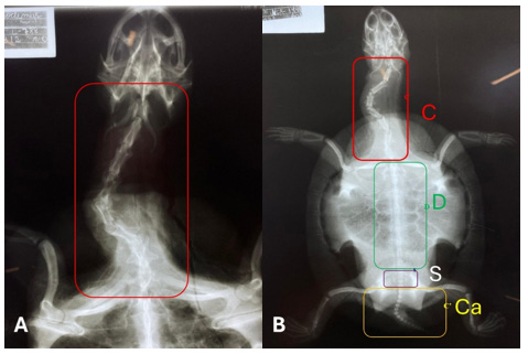
Regarding cervical vertebrae, five individuals presented seven vertebrae and five presented eight, with no association with sex (p > 0.05) or age. Dorsal vertebrae ranged between nine (n = 5) and ten (n = 5), also without correlation with the morphometric variables analyzed. The sacral region was constant, with three vertebrae in all specimens. The number of caudal vertebrae presented a mean of 18.0 ± 2.57 (range: 15–22). Stratified analysis by sex revealed no statistically significant differences in the total number of vertebrae between males and females, a finding consistent with previous reports in the literature (Mustafa et al., 2019). The variation in the number of cervical and dorsal vertebrae among specimens, with an even distribution between the observed configurations (n = 5 for each pattern), corroborates studies describing morphological plasticity in chelonians (Böhmer; Werneburg, 2017; Szczygielski, 2017).
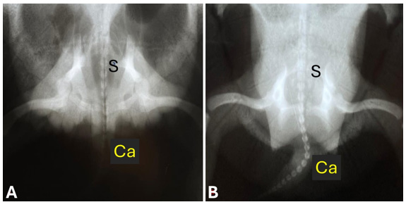
The cervical region (Figure 1), responsible for head retraction and mobility, exhibits greater anatomical flexibility, which may explain the differences found (Hermanson et al., 2022). In contrast, the dorsal region, structurally associated with support of the carapace and characterized by reduced mobility, showed less variability, while the sacral region remained constant in all specimens (Figure 2). Figure 1. Radiographic images in dorsoventral projection showing the cervical vertebrae (C) of Trachemys scripta elegans. In A: detail of the cervical region; in B: cervical (C), dorsal (D), sacral (S), and caudal (Ca) vertebrae. Scale bar: 1 cm. Figure 2. Radiographic images in dorsoventral projection showing the sacral (S) and caudal (C’) vertebrae of a female (A) and a male (B) Trachemys scripta elegans.
The tail is proportionally longer in the male. Scale bar: 1 cm. Observed was a positive and statistically significant correlation between body length and the number of caudal vertebrae (ρ = 0.764; p = 0.010; 95% CI = 0.27–0.94). However, T. s. elegans exhibits evident sexual dimorphism in the caudal region: although males present shorter body length compared to females, they possess proportionally longer and thicker tails, a characteristic associated with the presence of the copulatory organ (Gradela et al., 2017) (Figure 2). Thus, ontogenetic and sexual factors may act simultaneously in the variation of the caudal axis. However, considering the reduced sample size (n = 10) and the relatively wide confidence interval, studies with a larger number of individuals and sex-stratified analyses are necessary to confirm this tendency and determine the relative contribution of each factor.The positive correlation between body length and the number of caudal vertebrae indicates the influence of ontogenetic factors on axial development, with somatic growth associated with elongation and progressive differentiation of the caudal vertebrae (Duflot et al., 2024).
Radiography proved to be an effective tool for vertebral anatomical evaluation in chelonians, allowing identification of morphological patterns relevant for taxonomy, clinical practice, and comparative biology. The technique also contributes to management, conservation, and health assessment of wild animals maintained in captivity.
Conclusion
Radiography proved to be an effective method for anatomical characterization of the vertebral column of Trachemys scripta elegans. The vertebral formula showed variability in the cervical (7–8 vertebrae), dorsal (9–10), and caudal (mean 18 ± 2.57) regions, with a constant pattern of three sacral vertebrae. The positive correlation between body length and number of caudal vertebrae suggests the influence of ontogenetic factors on axial development. These data contribute to the comparative anatomy of chelonians and provide reference information for the clinical interpretation of radiographic examinations in the species. Future studies with larger samples and sex-stratified analyses are necessary to confirm the observed patterns. Acknowledgments The authors thank the National Council for Scientific and Technological Development (CNPq) for providing the scientific initiation scholarship.
Conflict of Interest
The authors declare that there is no conflict of interest related to the conduct, preparation, and publication of this article. Furthermore, they affirm that they did not receive any financial, personal, or institutional influences that could have interfered with the results or interpretations presented in this study. Source Of Funding This study was funded by the National Council for Scientific and Technological Development through the provision of a scientific initiation scholarship, which supported the development and execution of the research.
Figures and Tables
| Parameter | Mean (±DP) |
|---|---|
| Body length | 20,93 ± 1,79 cm |
| Carapace width | 16,21 ± 1,02 cm |
| Body height | 8,82 ± 0,99 cm |
| Body mass | 1.287,21 ± 226 g |
References
- BÖHMER, C.; WERNEBURG. I. Deep time perspective on turtle neck evolution: chasing the Hox code by vertebral morphology. Proceedings of the National Academy of Sciences, 114(51), 13677–13682, 2017.
- DUFLOT, B.; GILLET, A.; JONES, K. E.; SABIN, R.; LANZETTI, A. Ontogenetic changes in shape and growth rate during postnatal development in false killer whales (Pseudorca crassidens) vertebral column. Marine Mammal Science, 2024.
- GRADELA, A. et al. Sexual Dimorphism in Red-Eared Sliders (Trachemys scripta elegans) from the Wild Animal Triage Center of the Tiete Ecological Park, São Paulo, Brazil. Acta Scientiae Veterinariae, v. 45, p. 1468, 2017.
- HERMANSON, A. C. et al. Radiographic vertebral formula and anatomical variations in Trachemys dorbigni and Trachemys scripta elegans (Testudines: Emydidae). Anatomia Histologia Embryologia, [S. l.], v. 51, n. 3, p. 361–369, 2022. DOI: https://doi.org/10.1111/ahe.12736
- MAYERL, C. J.; BRAINERD, E. L.; BLOB, R. W. Pelvic girdle mobility of cryptodire and pleurodire turtles during walking and swimming. The Journal of Experimental Biology, v. 219, n. 17, p. 2650–2658, 2016.
- MUSTAFA, H.; LUTHFI, M. J.; ILMI, F.; KHOIRUNNISA, I.; TAKRIMA, T. Comparative anatomy of axial skeleton of red-eared turtle (TRACHEMYS SCRIPTA ELEGANS, Wied 1838) and softshell turtle (Amyda cartilaginea, Boddaert 1770). Proc. Int. Conf. Sci. Eng., v. 2, n. 1, p. 97-100, 2019.
- NOFITA, P.; LUTHFI, M. J.; DEWI, R. S. Anatomical study of red-eared turtle tail (Trachemys scripta elegans). Proc. Int. Conf. Sci. Eng., v. 2, n. 1, p. 57-62, 2021.
- SZCZYGIELSKI, T. Homeotic shift at the dawn of the turtle evolution. Roy. Soc. Open Sci., v. 4, p. 160933, 2017. Doi:10.1098/rsos.160933
- WERNEBURG, Ingmar; WILSON, Laura A. B.; PARR, William C. H.; JOYCE, Walter G. Evolution of neck vertebral shape and neck retraction at the transition to modern turtles: an integrated geometric morphometric approach. Systematic Biology, Oxford, v. 64, n. 2, p. 187– 204, 2015. DOI: 10.1093/sysbio/syu072
This is a text version generated from the article. For the formatted version of record (with original tables & figures), download the PDF →