Favorable long-term outcome of multiple brain lesions caused by candida albicans in a pediatric oncological patient
Rita Aldeia da Silva1 ✉
Rita Aldeia da SilvaCorresponding author- 1 Department of Pediatrics, Unidade Local de Saúde de Braga, Braga, Portugal
Cláudia P. Gonçalves1
Cláudia P. Gonçalves- 1 Department of Pediatrics, Unidade Local de Saúde de Braga, Braga, Portugal
Rafael Matias2
Rafael Matias- 2 Department of Radiotherapy, Instituto Português de Oncologia do Porto Francisco Gentil, Porto, Portugal
Ana Fraga3
Ana Fraga- 3 Department of Pediatrics Oncology, Instituto Português de Oncologia do Porto Francisco Gentil, Porto, Portugal
Filipa Almeida Leite3
Filipa Almeida Leite- 3 Department of Pediatrics Oncology, Instituto Português de Oncologia do Porto Francisco Gentil, Porto, Portugal
Show affiliations (3)
- 1Department of Pediatrics, Unidade Local de Saúde de Braga, Braga, Portugal
- 2Department of Radiotherapy, Instituto Português de Oncologia do Porto Francisco Gentil, Porto, Portugal
- 3Department of Pediatrics Oncology, Instituto Português de Oncologia do Porto Francisco Gentil, Porto, Portugal
Published
Abstract
We report the case of a 4-year-old girl with T-cell acute lymphoblastic leukemia who developed multiple brain abscesses caused by Candida albicans. Long-term antifungal therapy, tailored to the patient’s clinical course, led to disease regression, with the patient remaining asymptomatic after four years. This case highlights the importance of early diagnosis, individualized therapy, and meticulous follow-up in managing pediatric fungal central nervous system (CNS) infections.
1 Introduction#
Invasive fungal infections, particularly those involving the central nervous system (CNS), pose a significant challenge in pediatric oncology. Fungal brain abscesses in children undergoing chemotherapy remain uncommon but, despite significant advances in diagnostic and therapeutic strategies, are often associated with severe complications and poor outcomes. This report presents a case with an unexpectedly favorable outcome, emphasizing the importance of heightened clinical vigilance, early intervention, and tailored management strategies in such scenarios.
2 Case Report#
We describe the case of a 4-year-old girl diagnosed with T-cell acute lymphoblastic leukemia (ALL), who initially presented with fever and mediastinal lymph node enlargement. At the time of diagnosis, there was no evidence of CNS involvement: cerebrospinal fluid analysis was negative and initial magnetic resonance imaging (MRI) identified no abnormalities.
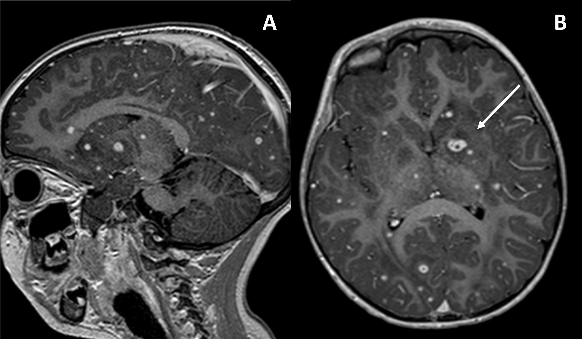
Three months into chemotherapy, the patient developed febrile neutropenia and acute respiratory failure secondary to fungal septicemia, with blood cultures positive for Candida albicans. A subsequent brain MRI showed multiple extensive lesions (Figure 1), leading to the initiation of Amphotericin B, Caspofungin, along with broad-spectrum antibiotics. Voriconazole was later added, leading to clinical improvement without neurological symptoms.
Although the patient initially exhibited clinical improvement, she was readmitted nine days later with neurological symptoms, including headache, nystagmus, and absence seizures. Suspecting Voriconazole toxicity, the drug was discontinued, and Levetiracetam was initiated.
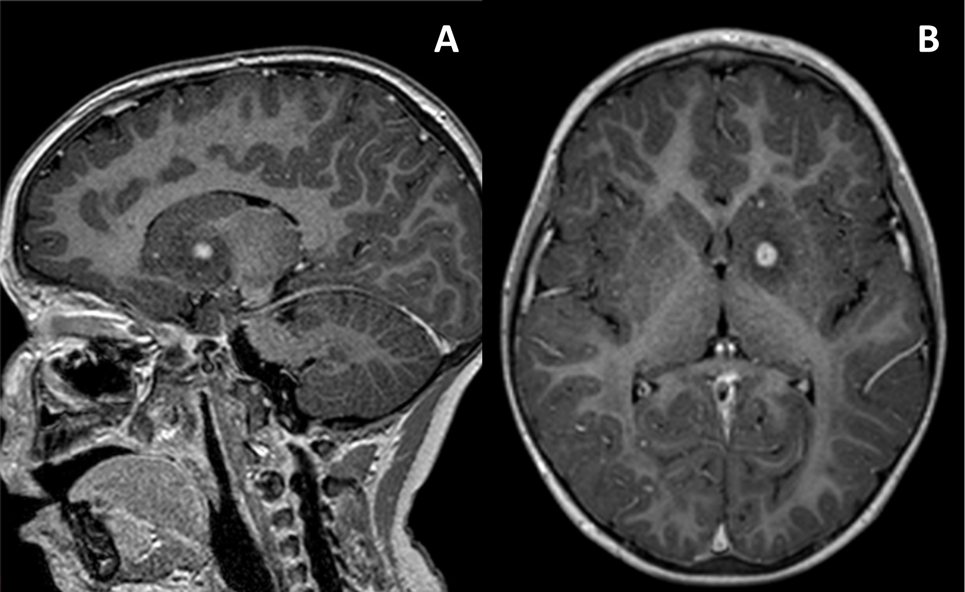
An MRI performed one year later revealed disease progression, prompting the initiation of Flucytosine, which led to disease regression. However, due to toxicity, it was subsequently discontinued. Five months later, despite the presence of multiple millimetric lesions and one larger stable lesion measuring 1 mm, the imaging confirmed a remarkable radiological response (Figure 2).
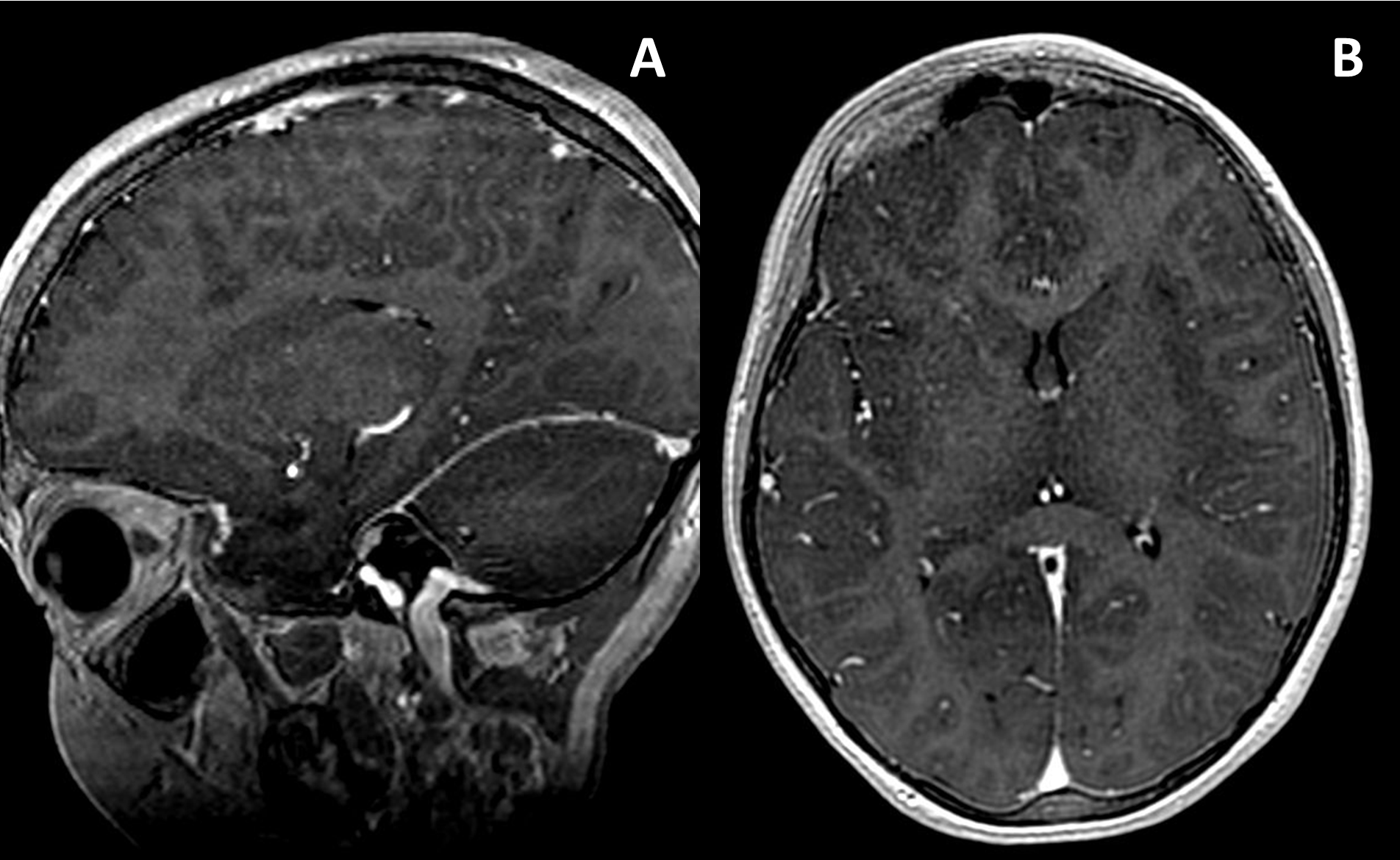
After four years of continuous antifungal therapy with Amphotericin B, the patient, now 8 years old, remained asymptomatic and demonstrated normal physical and neurological development. The most recent MRI (Figure 3) showed apparent resolution of the lesions, with no gadolinium-enhancing abnormalities.
3 Discussion#
Fungal brain abscesses represent rare yet severe complications in pediatric patients with hematologic malignancies (1). These infections present diverse clinical manifestations and are compounded by the aggressiveness of pathogens such as Candida and Aspergillus species, which are associated with high morbidity and mortality rates, emphasizing the importance of early diagnosis and rigorous therapeutic intervention (2,3). Despite aggressive antifungal therapies and surgical interventions, the literature highlights a significant risk of recurrence or progression, especially in severely immunocompromised patients (4).
In reviewed cases, mortality rates for Candida cerebral abscesses have reached nearly 70% (5). Other studies indicate that this high mortality is partly due to diagnostic challenges and delays in initiating appropriate therapy (6).
The diagnosis of Candida CNS infections remains challenging due to nonspecific clinical and radiological findings. Tissue biopsy is often necessary for diagnostic confirmation, as blood and cerebrospinal fluid (CSF) cultures are frequently nondiagnostic (5). Advanced imaging techniques, such as diffusion-weighted MRI, have improved the identification of fungal abscesses but cannot reliably differentiate them from bacterial or protozoal infections (5,7).
Management is equally complex due to the limited CNS penetration of many antifungal agents. Voriconazole, despite its efficacy and superior CNS penetration, is often limited by its significant toxicity profile, including hepatotoxicity, neurotoxicity, and visual disturbances, requiring careful monitoring during prolonged therapies (5). While the combination of voriconazole and Amphotericin B has shown efficacy in cases where surgical intervention is unfeasible (7), flucytosine offers an additional therapeutic option with excellent CNS penetration, serving as a critical adjunct to Amphotericin B in managing neurocandidiasis. However, flucytosine's use is limited by its risk of hematological toxicity, including leukopenia and thrombocytopenia, requiring close monitoring during therapy (6). These findings emphasize the importance of multidisciplinary collaboration and timely intervention to improve patient outcomes.
This report is particularly significant given the rarity of pediatric fungal brain abscesses. The patient’s prolonged therapy with Amphotericin B, combined with the sequential use of various antifungals, underscores the complexity of treatment strategies required for such infections. While the prognosis for fungal brain abscesses in pediatric patients is generally guarded, this case adds to the limited body of evidence, offering valuable insights into the potential for successful outcomes with carefully tailored, long-term management.
4 Conclusion#
The importance of maintaining a high clinical suspicion for fungal CNS involvement in pediatric ALL patients who develop new neurological symptoms during chemotherapy cannot be overstated. Early and aggressive intervention, combined with meticulous monitoring, is essential to mitigate severe consequences. This case underscores the potential for successful outcomes through sustained, individualized antifungal therapy and emphasizes the critical need for further research to enhance diagnostic and therapeutic strategies.
This case adds valuable data to the limited evidence available on pediatric fungal CNS infections, guiding future clinical practice.
4.1 Educational Objectives#
1. Early Recognition: High clinical suspicion is vital for diagnosing fungal CNS infections in immunocompromised children.
2. Evolving Treatment Strategies: Sequential, long-term antifungal therapy tailored to the patient’s clinical response can lead to positive outcomes.
3. Essential Monitoring: Rigorous follow-up with imaging and clinical assessment is crucial to detect disease progression and guide therapeutic adjustments.
Article information
- Published
Copyright © 2024 Rita Aldeia da Silva. This is an open access article distributed under the Creative Commons Attribution License, which permits unrestricted use, distribution, and reproduction in any medium, provided the original work is properly cited. CC BY 4.0 · Corrections policy
References 7#
- 1
Athanassiadou F, Tragiannidis A, Papageorgiou T, Velegraki A. Fungal brain abscesses in leukemia. Indian Pediatr. 2006;43(11):991–4.
- 2
Bilgin E, Ökten Aİ, Gezercan Y, et al. Pediatric Giant Cerebral Candida Abscess: A Case Report. Pediatr Neurosurg. 2019;54(3):207–11. doi:10.1159/000500678
- 3
Mehta A, Yadav M, Gupta BK, et al. Multiple brain abscesses in a neonate: a rare case report along with review of literature. Ann Med Surg (Lond). 2024;86(8):4793–8. Published 2024 May 17. doi:10.1097/MS9.0000000000002155
- 4
Frazier JL, Ahn ES, Jallo GI. Management of brain abscesses in children. Neurosurg Focus. 2008;24(6). doi:10.3171/FOC/2008/24/6/E8
- 5
Fennelly AM, Slenker AK, Murphy LC, Moussouttas M, DeSimone JA. Candida cerebral abscesses: a case report and review of the literature. Med Mycol. 2013;51(7):779-784. doi:10.3109/13693786.2013.789566
- 6
Sales MS, Leite MMB, Assunção RCG, Grangeiro LL. Candida species brain abscess: a life-threatening condition. Brazilian Journal of Health Review. 2022;5(3):9060-9076. doi:10.34119/bjhrv5n3-084.
- 7
Patiroglu T, Unal E, Karakukcu M, et al. Multiple fungal brain abscesses in a child with acute lymphoblastic leukemia. Mycopathologia. 2012;174(5-6):505-509. doi:10.1007/s11046-012-9563-6