Full Text Hide / show
Introduction
Non-Hodgkin lymphoma (NHL) refers to a diverse set of malignancies that originate from lymphoreticular tissue. It ranks as the second most prevalent cancer in the head and neck region, following squamous cell carcinoma (SCC). Lymph nodes are the primary sites affected by non-Hodgkin lymphoma (NHL); however, in certain Asian countries, the occurrence of this cancer in extranodal locations is notably higher. Additionally, there has been a rising trend in extranodal NHL cases in Western countries over recent decades. Approximately 23% to 30% of extranodal NHL cases, primarily involving B-cell NHL, mainly impact the gastrointestinal tract, skin, and bones. T-cell NHL is less prevalent and poses challenges in classification(1). Peripheral T-cell lymphoma, not otherwise specified (PTCL- NOS), is a subtype of T cell lymphoma that arises from the abnormal maturation of lymphoid lineage progenitor cells.
It is characterized by poorly defined features, which complicates its diagnosis.(2) While oral non-Hodgkin lymphoma (NHL) lesions typically result from widespread systemic involvement, rare instances may begin in the oral tissues before spreading elsewhere. Primary NHL manifestation in the mouth and lips is rare, accounting for approximately 2% of all NHL cases occurring outside of the lymph nodes.(1,2)The clinical appearance of oral non- Hodgkin lymphoma (NHL) varies, potentially manifesting as swelling, ulceration, or bone loss, particularly in areas like the gingiva and hard palate. Retrospective studies spanning the past 45 years indicate an exceptionally low occurrence of T-cell lymphoma (TCL) in the mouth, accounting for less than 3% of extranodal NHL cases with oral involvement.(1) This case report would describe the disease course and diagnostic dilemma while reaching the correct diagnosis of T-cell lymphoma of lip and would add up a learning point regarding a rare presentation of T-cell lymphoma.It also emphasizes the importance of adequate sampling needed for a correct diagnosis.
Case Description
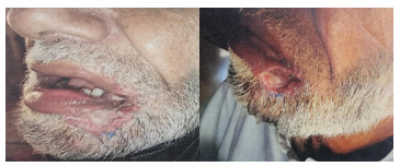
We present the case of 70 yrs old male with no known comorbidity presented in Oct’22 with history of left lower lip lesion for last few months gradually increasing in size not associated with fever,weight loss and night sweats. On examination there was an ulcerated area seen at left lower lip along with palpable left axillary and right inguinal lymphadenopathy while no hepatosplenomegaly appreciated. Fig.1. His past medical, past surgical, personal,drug and family history was unremarkable. Figure 1. An ulcerated area seen extending from left angle of mouth up to half of left lower lip. He underwent lip wound biopsy on 27-8-22 Immunohistochemistry showed positivity for CD3, weak positivity for CD30;Tdt,EMA and Alk were negative,concluded as partly ulcerated hyperplastic squamous epithelium with dense lymphoid infiltrates with no evidence of dysplasia or invasive squamous cell carcinoma.Second opinion taken for same biopsy from another laboratory on 12-9-22, immunohistochemistry positive for CD79a andCD20 in reactive B cells, MUM1 positive in plasm, CD3 highlights reactive T-cells,CD20 highlights reactive B cells.Ki67 30% proliferative index concluded as squamous mucosa with marked ulceration and necroinflammatory debris.
Because of non healing ulcer another incisional biopsy of cheek and left lower lip was done on 20-9-22 extended immunohistochemical profile were applied and concluded as atypical lymphoid infiltrate and surface covered by benign squamous epithelium with Ki67 of 70-80%.CT Neck Chest and Abdomen was done on 10-10-2022 that showed neoplastic lesion in lower lip and left buccal mucosa with subcentimeter bilateral neck lymph nodes along with bilateral axillary, subpectoral, right internal mammary, portocaval, iliac vessel and inguinal region lymph nodes suggestive of lymphoproliferative disorder. This is followed by left axillary lymph node biopsy on 13-10-22 showed reactive lymph node, negative for granuloma or malignancy.Again a second review of incisional biopsy (done on 20-9-22) was requested on 28-10-22 because of raised Ki-67 and *now favoured Peripheral T-cell lymphoma NOS with positivity for CD3 and CD56 with ki-67 of 50% while negative for Tdt, CD20 and EBV.
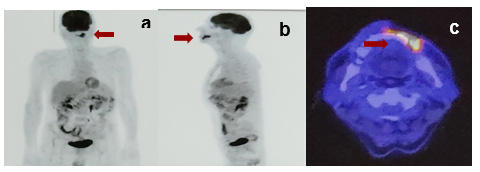
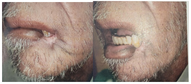
PET scan done on 29-10-22 showed FDG avid lesion in left lower lip and angle of mouth with SUVmax of 15.66 mea 4.5*1.5*2.8cm while reactive neck, portacaval and iliac vessels lymph nodes.Fig.2 Figure 2. Images (a)Coronal and (b) are Sagittal images from the PET SCAN showing an enhancing mass around oral cavity pointed with red arrow and image (c) showing an FDG avid lesion in left lower lip and angle of mouth with SUVmax of 15.66 mea 4.5*1.5*2.8cm. He was started on chemotherapy without waiting for the report with cycle 1 of Cyclophosphamide with 50% dose reduction,Vincristine and Prednisolone on 31-10-2022. Later on report came out on 4-11-2022 this time *giving a diagnosis of peripheral T-cell lymphoma , NOS is favoured showed CD3 and CD56 positive,CD20 Tdt and EBV negative with Ki-67 of 50%.The case was discussed in Tumor board meeting on 25-11-2022 and decided to continue with CHOP (Cyclophosphamide,Vincristine,Adriamycin and Prednisolone)for 4 more cycles followed by imaging.Patient received 3C of CHOP (Cyclophosphamide,Adriamycin,Vincristine and prednisolone) till 2-1-2023 followed by PET scan on 21-2-2023 showed complete response with DS 1.Fig.2 and 3 Figure 3.
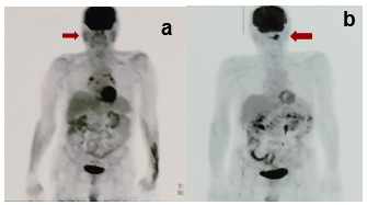
Complete resolution of lesion around Lf angle of mouth and lower lip along with visible fibrosis and granulation tissue seen. Figure 4. Compared to previous PET scan Oct,22 (b) No lesion seen on coronal and sagittal images of PET scan of Feb,23 (a) He was continued with 2 more cycles of CHOP x 6C till 17- 3-2023.PET scan repeated on 5-5-2023 showed sustained complete metabolic response.Case was re-discussed in Tumor board meeting and it was decided to get adjuvant radiation ; so received 5Fr from 27-5-2023 till 31-5-2023.PET scan then repeated post XRT on 23-8-2023 showed sustained metabolic response.He then remained on regular follow up till jan 2025
Discussion
We found that diagnosing peripheral T-cell lymphoma, not otherwise specified (PTCL-NOS), as a primary lesion in the lip posed significant challenges due to its rarity and atypical presentation. A similar case was reported by Asadi et al., who reported a 34-year-old male with an atypical presentation of PTCL-NOS as an ulcerated buccal mucosa lesion, a rare extranodal site ultimately confirmed through immunohistochemistry and successfully treated with chemotherapy and radiotherapy (19-3). The patient's nonhealing ulcer was initially misdiagnosed as benign, highlighting how these cases can mimic more common oral pathologies, highlighting how such cases can mimic more common oral pathologies. For instance, a case of primary diffuse large B-cell lymphoma (DLBCL) presented as a hyperplastic gingival lesion, leading to a delayed diagnosis until biopsy and immunohistochemistry confirmed the malignancy (Wiley, 2018).
This parallels our case, where the benign appearance of the lesion masked the underlying lymphoma, emphasizing the need for early tissue sampling and thorough pathological evaluation in atypical oral lesions. (20-4) The importance of early diagnosis cannot be overstated. Delayed diagnosis of aggressive subtypes like extranodal natural killer/T-cell lymphoma has been associated with poor outcomes, including significant tissue destruction. In our case, early diagnosis facilitated timely intervention with CHOP chemotherapy, leading to complete remission. The patient has remained disease-free for over two years, highlighting the benefits of prompt and accurate diagnosis.(21-5) Achieving an accurate diagnosis ultimately required multiple biopsies, second opinions, and an extended immunohistochemical panel, emphasizing the critical role of repeated tissue sampling.
Additionally, imaging, particularly PET scans, played a pivotal role in assessing disease extent and guiding management when histopathological findings remained inconclusive. In our case, the journey to an accurate diagnosis mirrors findings from comparative studies that emphasize the value of combining multiple diagnostic modalities. As reported in Blood, consensus diagnoses for PTCL subtypes were achieved only 74% to 81% of the time, even among expert pathologists. Similarly, our patient’s initial biopsies were inconclusive, necessitating repeated tissue sampling, second opinions, and an extended immunohistochemical panel to confirm PTCL-NOS. (22-6) Furthermore, the role of PET/CT in our case — not only for staging but also for guiding management and monitoring response, aligns with studies from the American Journal of Roentgenology and Frontiers in Oncology, which demonstrated PET/CT’s accuracy in both baseline staging and detecting disease involvement.
In our patient, PET/CT identified widespread lymphadenopathy despite negative lymph node biopsies, reinforcing how imaging can uncover disease burden when histology alone falls short.(23-7) The parallels between our case and these studies underscore the necessity of a multidisciplinary, multimodal approach — combining pathology, immunohistochemistry, molecular analysis, and advanced imaging — to overcome diagnostic uncertainty and optimize patient outcomes. The patient’s complete response to CHOP chemotherapy, followed by sustained metabolic remission, further supports the importance of timely treatment despite initial diagnostic delays. Treatment approaches for PTCL-NOS vary, but CHOP chemotherapy remains a common regimen. In a reported case, a 34-year-old male with PTCL-NOS of the buccal mucosa achieved disease-free status after receiving chemotherapy followed by radiotherapy, with no recurrence after 12 months.
Similarly, our patient responded well to CHOP chemotherapy, reinforcing the efficacy of this treatment modality. (19-3) This case highlights the importance of maintaining a high index of suspicion for lymphoma in persistent, unexplained oral lesions and demonstrates the value of thorough diagnostic workups and multidisciplinary management in improving outcomes.
Conclusion
This case underscores the importance of considering lymphoma as a differential diagnosis in persistent, nonhealing oral lesions, even in the absence of systemic symptoms. The diagnostic challenges associated with PTCL- NOS, particularly in extranodal sites like the lip, necessitate a thorough and multidisciplinary approach, including multiple biopsies, immunohistochemistry, and advanced imaging. Early recognition and timely initiation of chemotherapy, as in this case, can lead to successful remission and improved patient outcomes.. This case contributes to the growing body of literature on rare extranodal presentations of T-cell lymphomas and emphasizes the critical role of early diagnosis and individualized treatment strategies in optimizing prognosis.
Figures and Tables
References
- Dos Santos HL, Alves CG, Almendra Mattos RM, Curi DS, Leite Ribeiro P, Sarmento VA. Peripheral T-cell non- Hodgkin lymphoma manifesting as a primary lesion on the lip: a rare case report. Special Care in Dentistry. 2018 Nov;38(6):438-44.
- Vigouroux A, Lillo F, Hofer FD. Peripheral T-Cell Lymphoma not otherwise specified with oral manifestation: A Case Report. Journal of Oral Research. 2020;9(6):516-21.
- Asadi AA, Mahmoudi H, Mofidi A, Mortezazadeh M, Hadizadeh A. Peripheral T-cell lymphoma of the oral cavity: A case report. Cancer Reports. 2023 Jan;6(1):e1751.
- Donaduzzi LC, Reinheimer A, da Silva MA, de Noronha L, Johann AC, Franco A, Couto SD, Souza PH. Primary diffuse large B cell lymphoma mimicking hyperplastic reactive lesion (lymphoma of the oral cavity). Case reports in pathology. 2018;2018(1):2981689.
- Schuler A, Smith E, Lowe L, Helfrich Y. Extranodal natural killer/T-cell lymphoma, nasal type: a rare but critical diagnosis. JAAD case reports. 2017 May 1;3(3):225-7.
- Moskowitz AJ, Lunning MA, Horwitz SM. How I treat the peripheral T-cell lymphomas. Blood, The Journal of the American Society of Hematology. 2014 Apr 24;123(17):2636-44.
- Cronin CG, Swords R, Truong MT, Viswanathan C, Rohren E, Giles FJ, O'Dwyer M, Bruzzi JF. Clinical utility of PET/CT in lymphoma. American Journal of Roentgenology. 2010 Jan;194(1):W91-103.
- de Oliveira EM, de Cáceres CV, Fernandes-Rodrigues CI, Penafort PV, Legarrea JM, Gomes NR, Pontes HA, Vargas PA, Júnior JN, Soares CD, Fonseca FP. Oral manifestations of peripheral T cell lymphoma, not otherwise specified: case series and review of the current literature. Oral Surgery, Oral Medicine, Oral Pathology and Oral Radiology. 2024 Aug 17.
- Yang H, Qiu M, Zhou Y, Jiang L, Dan H, Chen Q. NK/T- Cell Lymphoma on the Upper Lip. J Mol Biomark Diagn. 2016;7(288):2.
- Kaplan I, Shuster A, Frenkel G, Avishai G, Allon I, Raiser V. Non-Hodgkin lymphoma of the lips: a rare entity. Acta Histochemica. 2019 Nov 1;121(8):151449.
- Villa A, Mariani U, Villa F. T-cell lymphoma of the oral cavity: a case report. Australian dental journal. 2010 Jun;55(2):203-6.
- Yin HF, Jamlikhanova V, Okada N, Takagi M. Primary natural killer/T-cell lymphomas of the oral cavity are aggressive neoplasms. Virchows Archiv. 1999 Oct;435:400-6.
- Miller RI. Non-Hodgkin's lymphoma of the lip: A case report. Journal of oral and maxillofacial surgery. 1993 Apr 1;51(4):420-2.
- Chandu A, Mitchell DA, Corrigan AM. Cutaneous CD30 positive large T cell lymphoma of the upper lip: a rare presentation. British Journal of Oral and Maxillofacial Surgery. 1997 Jun 1;35(3):193-5.
- Silva TD, Ferreira CB, Leite GB, de Menezes Pontes JR, Antunes HS. Oral manifestations of lymphoma: a systematic review. Ecancermedicalscience. 2016;10.
- Travassos DC, Silveira HA, Silva EV, Panucci BZ, da Silva Filho NC, Silva PV, Bufalino A, León JE. Primary cutaneous CD8+ cytotoxic T-cell lymphoma of the face with intraoral involvement, resulting in facial nerve palsy after chemotherapy. Journal of Cutaneous Pathology. 2022 Jun;49(6):560-4.
- Park JH, Shin HT, Lee DY, Ko YH. Extranodal natural killer (NK)/T-cell lymphoma presenting with recurrent lip swelling. International Journal of Dermatology. 2013 Jun 1;52(6).
- Chim CS, Chan AC, Raymond L. Primary CD30-positive anaplastic large cell lymphoma of the lip. Oral oncology. 1998 Jul 1;34(4):313-5.
- Deng D, Wang Y, Liu W, Qian Y. Oral and maxillofacial non-Hodgkin lymphomas: Case report with review of literature. Medicine. 2017 Sep 1;96(35):e7890..
- Scully C, Eveson JW, Witherow H, Young AH, Tan RS, Gilby ED. Oral presentation of lymphoma: case report of T-cell lymphoma masquerading as oral Crohn's disease, and review of the literature. European Journal of Cancer Part B: Oral Oncology. 1993 Jan 1;29(3):225-9.
- Khan UJ, Alamgir W, Ashraf A, Haider A, Rashid S, Abrar F. CLINICOPATHOLOGICALANDIMMUNOHISTOCHEMICAL PROFILE OF LYMPHOMA OF HEAD AND NECK REGION. Pakistan Journal of Pathology. 2021 Dec 31;32(4):142-6.
- Miyagawa F, Ogawa K, Asada H. A case of CD4+/CD8+ double-positive primary cutaneous anaplastic large cell lymphoma of the lip involving spontaneous regression after biopsy. European Journal of Dermatology. 2017 Jan 1;27(1):68-9.
- Tseng CH, Wang WC, Chen CY, Hsu HJ, Chen YK. Clinical manifestations of oral lymphomas–retrospective study of 15 cases in a Taiwanese population and a review of 592 cases from the literature. Journal of the Formosan Medical Association. 2021 Jan 1;120(1):361-70.
- de Figueiredo RH, Parreira BS, Canao PA, Cardoso L, Fonseca E, Almeida J. Peripheral T-Cell Lymphoma, Not Otherwise Specified–a case report and short literature review. Archive of Clinical Cases. 2022 Dec 19;9(4):140.
This is a text version generated from the article. For the formatted version of record (with original tables & figures), download the PDF →