Spontaneous cholecysto-cutaneous fistula
Meryam Mesbahi
Meryam Mesbahi More articles by this author →Haithem Zaafouri ✉
Haithem ZaafouriCorresponding author More articles by this author →Nizar Khedhiri
Nizar Khedhiri More articles by this author →Anis Ben Maamer
Anis Ben Maamer More articles by this author →
Published
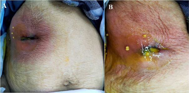
An 87-year-old female patient presented to our surgical department with complaints of persistent right upper abdominal pain lasting for three months and an inflammatory subcostal mass on the right side. The patient had a history of acute pancreatitis four months prior. On abdominal examination, a 6 cm abscess was noted in the right subcostal area, accompanied by cellulitis and discharge of bile and gallstones (Figure 1 Panel A and B). Laboratory results showed an elevated white blood cell count of 17000/mm3 and a CRP level of 170mg/dl, while other parameters were within normal limits.
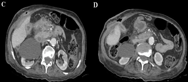
Due to local pain at the abscess site, abdominal ultrasound was not feasible. However, abdominal computed tomography revealed acute phlegmonous cholecystitis with a fistula extending to the skin, complicated by localized biliary peritonitis (Figure 2 Panel C and D). A diagnosis of necrotizing fasciitis involving the anterior abdominal wall and a cholecystocutaneous fistula was established.
The patient underwent laparotomy, confirming the imaging findings. An infected bile and digestive fluid effusion was observed, along with a gangrenous gallbladder fistula releasing calculi. Additionally, an abscess formed by the gallbladder. The inflamed gallbladder was freed from adhesions to the abdominal wall abscess, and multiple gallstones were removed from the subcutaneous tissue. Cholecystectomy was performed, followed by drainage of the subcutaneous abscess before abdominal closure. The postoperative course was uneventful.
Article information
- Published
Copyright © 2025 Dr. Haithem Zaafouri. This is an open access article distributed under the Creative Commons Attribution License, which permits unrestricted use, distribution, and reproduction in any medium, provided the original work is properly cited. CC BY 4.0 · Corrections policy