Full Text Hide / show
Background
Ulnar collateral ligament (UCL) injuries are clinically relevant in sports medicine because they may compromise joint stability, grip strength, pinch function, athletic performance, and return to sport. Although UCL injuries are frequently discussed in relation to the elbow, thumb metacarpophalangeal UCL injuries are particularly important in athletes involved in skiing, football, martial arts, wrestling, ball-handling sports, and contact activities. Objective: This systematic review aimed to compare surgical and conservative treatment for UCL rupture in athletes, with emphasis on return to sport, time to return, pain, function, joint stability, performance after return, and complications. Methodology: A systematic review was conducted according to PRISMA recommendations. Searches were performed in PubMed, MEDLINE, LILACS, and ScienceDirect for studies published between January 2010 and April 2026.
Eligible studies included athletes or sport-related populations with UCL injury, primarily involving the thumb metacarpophalangeal joint, and reporting surgical, conservative, diagnostic, functional, or return-to-sport outcomes. A total of 16 studies were included in the final qualitative synthesis. Data were synthesized qualitatively because of heterogeneity in anatomical site, study design, treatment strategy, rehabilitation protocols, and outcome definitions. Results: The included evidence was predominantly composed of retrospective cohorts, diagnostic studies, technical reports, case series, and case reports. Surgical treatment showed favorable outcomes in complete, unstable, displaced, chronic, bilateral, or high-demand thumb UCL injuries, with high rates of joint stability, functional recovery, and return to sport. Conservative treatment appeared appropriate for stable partial injuries when joint stability was preserved and no displacement or Stener lesion was suspected.
Diagnostic studies emphasized that clinical examination remains central to decision-making, while MRI, 3T MRI, and ultrasound may support diagnosis in equivocal or complex cases. Reported complications included stiffness, residual pain, neuroma, adhesions, tenolysis, overtensioning in augmentation procedures, and adverse events related to accelerated return-to-play protocols. Conclusion: Surgical treatment appears to provide more predictable restoration of stability and return-to-sport outcomes in complete or unstable thumb UCL injuries in athletes, whereas conservative management remains appropriate for stable partial lesions. Current evidence is limited by heterogeneous designs, small samples, and few direct comparisons between operative and nonoperative strategies. Future prospective studies should standardize injury classification, rehabilitation protocols, and return-to-sport criteria. Keywords: Athletic Injuries; Athletes; Thumb; Ulnar Collateral Ligament; Return to Sport.
Introduction
Ulnar collateral ligament (UCL) injuries represent a clinically relevant condition in sports medicine because they affect joints that are essential for athletic performance and return to sport. These injuries occur in anatomical regions exposed to valgus stress, axial loading, repetitive overload, and acute traumatic mechanisms. Although UCL injury is frequently associated with the elbow in overhead athletes, particularly baseball and softball players, it is also highly relevant to the thumb metacarpophalangeal joint, where ligament integrity is necessary for gripping, pinching, grasping, ball control, blocking, tackling, and contact-related athletic activities [1–3]. At the elbow, the UCL acts as the primary restraint against valgus stress during throwing, especially in the late cocking and early acceleration phases.
Repetitive overload may lead to progressive ligament attenuation, medial elbow pain, instability, loss of throwing velocity, reduced accuracy, and impaired competitive performance. These deficits directly affect the athlete’s ability to return to sport at the preinjury level. Although chronic attritional elbow UCL injury is commonly described in overhead throwers, acute traumatic rupture may also occur in athletes from other sports and may require different management depending on tissue quality, lesion pattern, and sport-specific demand [4,5]. At the thumb metacarpophalangeal joint, the UCL is essential for stability during pinch, grasp, opposition, and sport-specific manual tasks. Acute thumb UCL rupture is classically described as “skier’s thumb,” whereas chronic or attritional injury is known as “gamekeeper’s thumb.” The usual mechanism involves forced hyperabduction or hyperextension of the thumb, which may occur during skiing, football, martial arts, wrestling, ball-handling sports, or falls onto an abducted thumb [6–8].
In athletes, this injury may compromise grip strength, equipment control, ball handling, blocking capacity, and the ability to tolerate contact, all of which are fundamental for safe and effective return to sport. The clinical spectrum of UCL injury ranges from partial sprains with preserved stability to complete ruptures with marked laxity. In thumb injuries, complete rupture may be complicated by a Stener lesion, in which the torn ligament is displaced superficial to the adductor aponeurosis, preventing anatomical healing and increasing the risk of chronic instability, pain, weakness, and impaired function [9,10]. These consequences are particularly important in athletes because residual instability may delay returning to sport, reduce performance after return, or increase the risk of reinjury.
Treatment selection is guided by anatomical site, injury severity, degree of instability, tissue quality, presence of displacement, sport modality, competitive level, timing within the season, and functional demand. Stable partial injuries are usually managed nonoperatively with immobilization, bracing, and progressive rehabilitation, aiming to restore pain-free motion, strength, stability, and sport-specific function. In contrast, complete ruptures, displaced avulsion fractures, Stener lesions, chronic instability, failed conservative treatment, and high-demand athletic injuries are more frequently treated surgically through repair, reconstruction, or augmentation techniques to obtain more reliable joint stability and facilitate return to sport [11–16]. Return to sport is a central outcome in athletic populations and should not be limited to simple participation.
Ideally, the athlete should return safely, without pain or instability, and at a level comparable to preinjury performance. Operative treatment may provide more predictable restoration of stability in complete or unstable injuries, potentially supporting a more reliable return to sport. However, surgery may be associated with stiffness, residual pain, nerve irritation, implant-related symptoms, and complications related to accelerated return-to-play protocols. Conversely, conservative treatment avoids surgical morbidity and may be effective for stable partial injuries, but it may be insufficient for complete unstable ruptures, potentially resulting in persistent pain, weakness, residual instability, delayed return to sport, or inability to return to the previous competitive level [17–21]. Despite increasing interest in UCL injuries among athletes, the comparative effectiveness of operative and nonoperative treatment remains incompletely defined.
The available evidence is heterogeneous regarding anatomical sites, sport type, injury classification, treatment technique, rehabilitation protocol, and criteria used to define return to sport. Therefore, a systematic synthesis is necessary to clarify which treatment strategy offers the most favorable conditions for safe and effective return to sport after UCL rupture. The objective of this systematic review was to compare surgical and conservative treatment for UCL rupture in athletes, with emphasis on return to sport, time to return, pain, function, joint stability, performance after return, and complications. The hypothesis of this review is that surgical treatment provides more predictable joint stability, functional recovery, and return-to-sport outcomes than conservative treatment in athletes with complete, unstable, displaced, or high-demand UCL ruptures, whereas conservative management remains effective for partial and stable injuries.
Methodology
Study Design This study was conducted as a systematic review structured according to the Preferred Reporting Items for Systematic Reviews and Meta-Analyses (PRISMA) recommendations. The review was designed to compare operative and non-operative treatmentstrategiesforulnarcollateralligament(UCL)rupture in athletes, with an emphasis on clinical outcomes, functional recovery, and return to sport. Given the heterogeneity among the included studies regarding anatomical site, study design, injury pattern, treatment technique, rehabilitation protocol, and outcome measures, the data were synthesized primarily through a qualitative and narrative approach. This review is registered with PROSPERO under the registration number CRD42024658122. Research Question The review was guided by the following research question: in athletes with UCL rupture, does surgical treatment provide better clinical, functional, and return-to-sport outcomes than conservative treatment?
PICO Strategy
The research question was structured using the PICO framework. The population consisted of athletes with UCL rupture, including injuries involving both the elbow and the thumb metacarpophalangeal joint. The intervention was surgical treatment, including primary repair, suture anchor repair, subperiosteal repair, transosseous repair, ligament reconstruction with graft, internal brace augmentation, and suture tape augmentation. The comparison was conservative treatment, including immobilization, bracing, thumb spica casting, physical therapy, rehabilitation, and nonoperative return-to-play strategies. The outcomes of interest were return to sport, time to return to sport, recovery time, pain, range of motion, strength, patient-reported function, joint stability, complications, recurrence, and performance after return. Data Sources The search was performed in four electronic databases: PubMed, MEDLINE, LILACS, and ScienceDirect.
These databases were selected because they include biomedical, orthopedic, rehabilitation, and sports medicine literature relevant to UCL injuries. Search Period The search period was defined from January 2010 to April 2026. This interval was selected to include contemporary evidence regarding diagnostic methods, surgical techniques, conservative protocols, rehabilitation approaches, and return-to-sport criteria. Search Strategy The search strategy was developed to identify studies addressing ulnar collateral ligament injury in athletic populations, with emphasis on treatment strategy, rehabilitation, functional outcomes, and return to sport. The search combined diagnostic terms such as “Ulnar Collateral Ligament,” “UCL injury,” “UCL rupture,” and “UCL tear” with population-related terms, including “athletes,” “sports,” “baseball,” “overhead sports,” “football,” “skiing,” and “ballhandling sports.” Treatment-related terms included “surgery,” “surgical treatment,” “repair,” “reconstruction,” “conservative treatment,” “nonoperative treatment,” and “rehabilitation.” Outcome-related terms included “return to sport,” “return to play,”“functionaloutcomes,”“clinicaloutcomes,”and“recovery.” For PubMed, MEDLINE, ScienceDirect, and LILACS, Boolean operators were used to combine diagnostic, population, intervention, comparison, and outcome terms.
Both freetext terms and indexed terminology were considered when available, including expressions related to ulnar collateral ligament injuries. Although LILACS was included as a database source, only articles published in English were eligible for inclusion in the final review. Therefore, Portuguese descriptors were not used as inclusion criteria, and non- English publications were excluded during screening or fulltext eligibility assessment. The search terms were adapted according to the indexing structure and search interface of each database, while maintaining the same conceptual strategy across all sources. This approach was intended to maximize the identification of relevant English-language studies on operative and nonoperative management of UCL rupture in athletes. Inclusion Criteria Studies were considered eligible when they met the following criteria: publication between 2010 and 2026; inclusion of athletes, sports-related populations, or patients with sportrelated UCL injury; evaluation of thumb or elbow UCL injury; assessment of surgical treatment, conservative treatment, rehabilitation, or return-to-sport outcomes; reporting of clinical, functional, diagnostic, biomechanical, therapeutic, or return-to-sport data relevant to UCL management.
Clinical studies, prospective or retrospective cohorts, observational studies, case series, diagnostic studies, technical notes, systematic reviews with relevant contextual information, and case reports with direct clinical applicability were considered for qualitative synthesis when they contributed to the research question. Exclusion Criteria Studies were excluded when they did not address UCL injury or rupture, did not involve athletic or sport-related populations, lacked clinically relevant treatment or outcome data, focused exclusively on unrelated anatomical structures, presented duplicate or overlapping data, or consisted of editorials, commentaries, or publications with insufficient methodological detail. Articles that did not provide direct comparative data between surgical and conservative treatment but contributed relevant information regarding anatomy, imaging, diagnosis, injury mechanism, treatment indication, or return-to-sport decisionmaking were retained as supportingreferences.
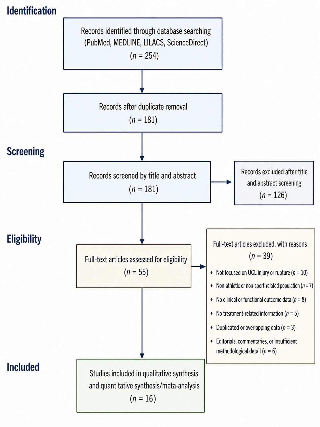
This approach was adopted because the available literature on UCL rupture in athletes is heterogeneous and includes studies that are clinically relevant even when not directly comparative. Study Selection Process The study selection process followed sequential PRISMA stages. First, all records identified in the databases were compiled. Duplicate records were removed. Second, titles and abstracts were screened to exclude studies clearly unrelated to UCL injury, athletic populations, treatment, or clinical outcomes. Third, potentially eligible articles underwent full-text assessment. During full-text review, studies were evaluated according to the inclusion and exclusion criteria and classified according to anatomical site, study design, population,intervention,comparator,andoutcomerelevance. Figure 1 presents the study selection process according to the PRISMA framework.
A total of 254 records were initially identified through searches in PubMed, MEDLINE, LILACS, and ScienceDirect. After duplicate removal, 181 records remained and were screened by title and abstract, resulting in the exclusion of 126 studies. Subsequently, 55 full-text articles were assessed for eligibility, of which 33 were excluded for not meeting the predefined inclusion criteria. Finally, 16 studies were included in qualitative synthesis and quantitative synthesis/meta-analysis. Insert Figure 1 here. Figure 1. PRISMA flow diagram of the study selection proces Data Extraction Dataextractionwasperformedusingastandardizedframework developed according to the review question and PICO strategy. The extracted variables included author, year of publication, anatomical site, study design, sample size, population characteristics, sport modality, injury type, treatment strategy, surgical technique, conservative protocol, rehabilitation approach, return-to-sport rate, time to return to sport, pain outcomes, range of motion, strength, patient-reported functional scores, joint stability, complications, recurrence, performance after return, and methodological limitations.
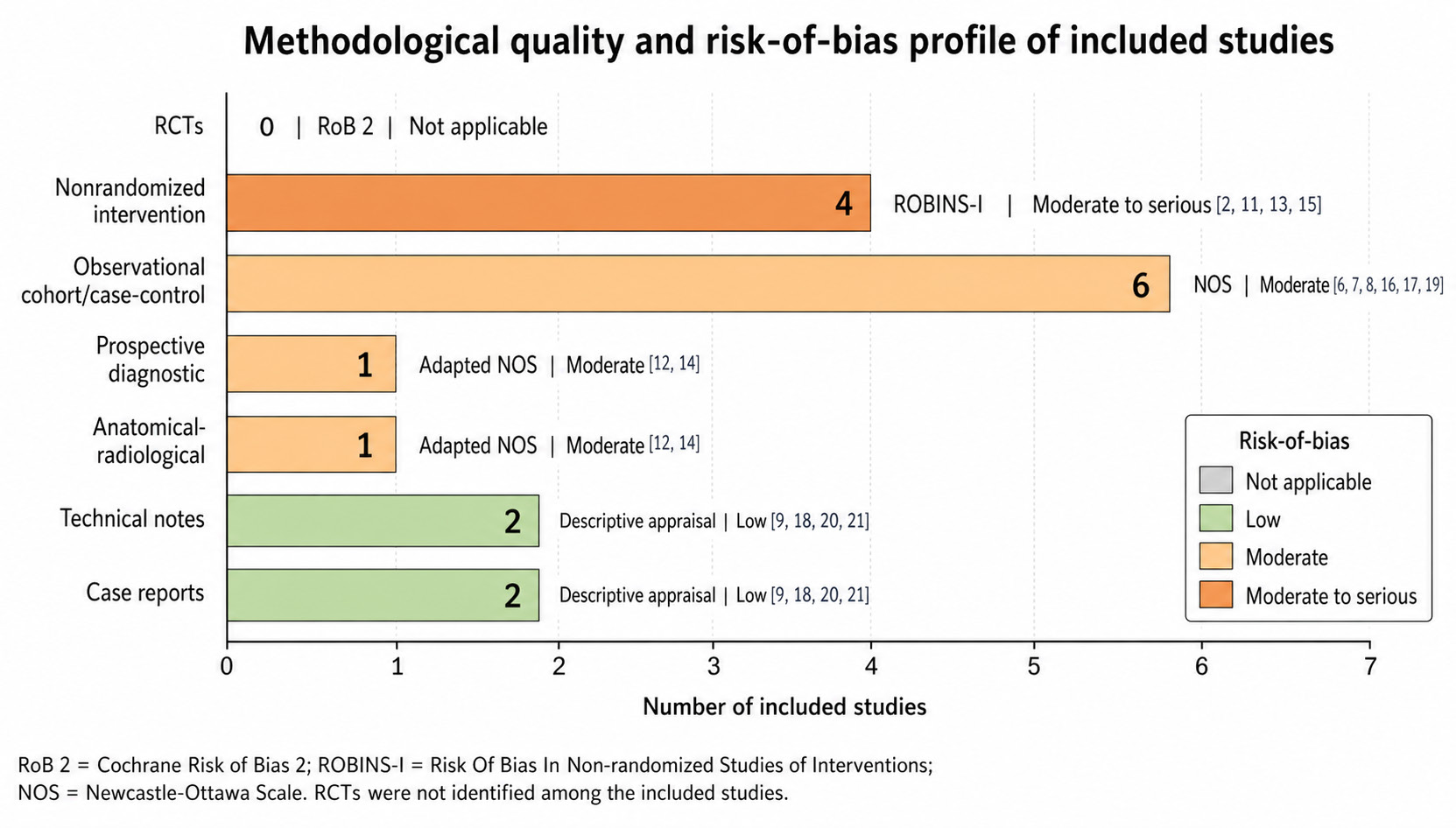
When return-to-sport data were reported, attention was given towhetherthestudydefinedreturnasanyparticipation,return to training, return to competition, or return to the preinjury level of performance. When definitions were unclear or inconsistent, this was recorded as a methodological limitation. Methodological Quality Assessment The methodological quality of observational studies was assessed conceptually using the Newcastle-Ottawa Scale framework, considering participant selection, comparability of groups, exposure or intervention assessment, outcome measurement, and adequacy of follow-up. Case reports and technical notes were evaluated descriptively according to diagnostic clarity, treatment description, clinical relevance, follow-up duration, and applicability to the review question. Theoverallqualityofevidencewasconsideredlowtomoderate because the included literature was predominantly composed of retrospective studies, case series, diagnostic studies, technicalnotes,biomechanicaloranatomicalreviews,andcase reports. The scarcity of randomized controlled trials and direct comparative studies between operative and nonoperative management limited the strength of causal conclusions.
Results
The results section presents the evidence extracted from the primary studies included in the review, with emphasis on study design, anatomical site, population characteristics, sportrelated context, treatment approach, diagnostic assessment, functional outcomes, and return-to-sport relevance. Because the included articles differed substantially in methodology and objectives, the findings were synthesized descriptively rather than through direct statistical pooling. Table 1 summarizes the general characteristics of the 16 primary studies included in the synthesis, organized according to study category, anatomical region, population profile, and main research focus. The included evidence was predominantly centered on injuries of the thumb metacarpophalangeal joint, especially ulnar collateral ligament lesions, although some studies also addressed lesser metacarpophalangeal joints, interphalangeal injuries, and broader sports-related hand trauma.
The table also demonstrates the methodological heterogeneity of the included literature. Surgical outcome studies represented the largest group, focusing on repair, reconstruction, augmentation techniques, functional recovery, stability, and return to sport. Diagnostic and imaging studies contributed evidence on clinical examination, MRI, 3T MRI, ultrasound, and imaging-based instability assessment, while anatomical, morphological, technical, and case-based studies provided complementary information regarding injury mechanisms, surgical decision-making, diagnostic pitfalls, and rare clinical presentations. Table 1. General characteristics of the included studies. Study category Included studies Anatomical region Population profile Main focus Surgical outcome studies Ly et al., 2023; Legerstee et al., 2025; Potter et al., 2025; Sochacki et al., 2018; Najd Mazhar et al., 2025 Predominantly thumb MCP joint Mixed populations, high-level athletes, professional football players, and individual case reports Clinical outcomes, functional recovery, stability, and return to sport after surgical repair or reconstruction Diagnostic and imaging studies Mahajan et al., 2016; Bai et al., 2018; Orbenes et al., 2025; Goodarzi et al., 2025; Hoy et al., 2026 Thumb MCP joint, fingers, and thumb Mixed or sports-related populations Diagnostic accuracy of clinical examination, MRI, 3T MRI, ultrasound, and imagingbased instability assessment Anatomical and morphological studies Legerstee et al., 2024; Vicentini et al., 2026 Thumb UCL avulsion and lesser MCP joints General or mixed populations Avulsion morphology, collateral ligament injury patterns, and associated softtissue injuries Technical reports and case-based evidence Arvind & Strauch, 2025; Kress et al., 2025; Bernstein et al., 2020; Amiel et al., 2025 Thumb MCP or IP region General population, collegiate athletes, and recreational athletes Description of surgical techniques, protected return to play, diagnostic pitfalls, and rare injury presentations Legend: MCP = metacarpophalangeal; IP = interphalangeal; UCL = ulnar collateral ligament; RCL = radial collateral ligament; MRI = magneticresonanceimaging;3TMRI=3-Teslamagneticresonance imaging; NFL = National Football League; NCAA = National CollegiateAthleticAssociation;N/A=notapplicableornotreported.
Return to sport was most consistently reported in studies involving athletic populations, although the definition of this outcome varied across the included articles. Some studies defined return to sport as return to recreational activity or daily sport participation, whereas others considered return to the preinjury competitive level. Overall, surgical studies provided the most consistent return-to-sport data, particularly in athletes with complete, unstable, or high-demand thumb UCL injuries. Sochacki et al. (2018) reported that 22 of 23 National Football League players returned to sport after thumb UCL surgery, corresponding to a 95.7% return-to-sport rate, with a mean time to return of 132.2 days [16]. Bernstein et al. (2020) reported return to play in three collegiate football linemen at a mean of 13.3 days after thumb UCL repair with protective thumb spica gauntlet casting; however, this accelerated strategy was associated with adjacent joint dislocations [15].
Potter et al. (2025) reported return to sport or activity between 8 and 12 weeks after collateral ligament repair using a bio-composite scaffold in a predominantly athletic cohort [13]. Najd Mazhar et al. (2025) described complete recovery of mobility, strength, and stability at six months after bilateral surgical repair in a Taekwondo athlete [21]. The only conservatively managed athlete-specific case was reported by Amiel et al. (2025), in which a recreational Newcomb ball player returned to full activity after diagnostic correction and rehabilitation at 12 weeks [20]. Functional recovery and restoration of joint stability were also favorable in most surgically treated cohorts. Ly et al. (2023) reported excellent stability in 91% to 100% of surgically treated grade III thumb UCL injuries, with low postoperative disability scores, including a mean QuickDASH of 8.7/100 and PRWE of 7.1/100 [2].
Legerstee et al. (2025) reported improved patientreported outcomes after tendon autograft reconstruction, with 84% of patients regaining metacarpophalangeal stability [11]. Potter et al. (2025) observed marked functional improvement, with QuickDASH decreasing from 81.5 to 4.1 after repair or augmentation using a bio-composite scaffold [13]. Arvind and Strauch (2025) reported improvement in thumb range of motion from 20° to 45° after anchorless repair, although this evidence was limited to a single case [9]. Complications varied according to injury severity, surgical technique, and rehabilitation strategy. Surgical cohorts reported stiffness, residual pain, neuroma, adhesions, tenolysis, transient inflammatory reactions, and technical risks such as overtensioning in augmentation procedures [2,11,13,18]. Early return-to-play protocols also presented specific concerns, as Bernstein et al.
(2020) reported adjacent joint dislocations during protected return with thumb spica gauntlet casting [15]. In conservatively managed or initially misdiagnosed cases, complications were mainly related to unnecessary immobilization, stiffness, or delayed diagnostic correction [20]. Table 2 summarizes the main clinical outcomes, including return-to-sport results, functional recovery, time to return, and reported complications. This table was constructed to integrate the key outcome domains into a single synthesis, allowing comparison across surgical techniques, conservative rehabilitation, and case-based evidence. It highlights that surgical treatment was associated with high return-tosport rates and favorable functional outcomes, whereas conservative evidence was limited to selected or case-based contexts. Table 2. Clinical, functional, return-to-sport, and complication outcomes. Author/Year Treatment or Context Return-to-Sport / Functional Outcome Time to Return Complications / Limitations Ly et al., 2023 [2] Surgical treatment of grade III thumb UCL injuries Excellent stability in 91–100%; QuickDASH 8.7/100; PRWE 7.1/100 Mean 2.9 months Stiffness 17%; residual pain 26% Arvind and Strauch, 2025 [9] Anchorless thumb UCL repair ROM improved from 20° to 45° Not reported Single case; limited external validity Legerstee et al., 2025 [11] Tendon autograft reconstruction Improved patientreported outcomes; 84% regained MCP stability Not reported Neuroma, adhesions, tenolysis Potter et al., 2025 [13] Bio-composite scaffold repair/ augmentation QuickDASH improved from 81.5 to 4.1; return to activity reported 8–12 weeks Mild transient erythematous reaction; small cohort Bernstein et al., 2020 [15] Surgical repair with thumb spica gauntlet casting Full return to preinjury level in collegiate football linemen Mean 13.3 days Adjacent joint dislocations Sochacki et al., 2018 [16] Thumb UCL surgery in NFL players 95.7% returned to sport; no overall significant performance decline Mean 132.2 days Position-specific performance decline reported Kress et al., 2025 [18] Suture tape augmentation Higher construct strength; supports accelerated rehabilitation Potential 5–6 weeks Risk of overtensioning; technical evidence Amiel et al., 2025 [20] Conservative rehabilitation after diagnostic correction Full recovery of activity 12 weeks Stiffness after unnecessary initial immobilization Najd Mazhar et al., 2025 [21] Bilateral surgical repair Full recovery of mobility, strength, and stability 6 months Single case; no major complication reported Legend: UCL = ulnar collateral ligament; MCP = metacarpophalangeal; QuickDASH = shortened Disabilities of the Arm, Shoulder and Hand questionnaire; PRWE = Patient-Rated Wrist Evaluation; MHQ = Michigan Hand Outcomes Questionnaire; ROM = range of motion; NFL = National Football League.
Diagnostic accuracy was an important component of the included evidence because treatment selection depends on the ability to distinguish partial from complete ruptures, identify instability, detect avulsion fractures, and recognize Stener lesions. Mahajan et al. (2016) showed that careful clinical examination may reduce the need for MRI in suspected skier’s thumb, particularly when the diagnosis is clinically evident [12]. Orbenes et al. (2025) found that MRI may demonstrate high diagnostic sensitivity but does not necessarily change surgical decision-making beyond careful physical examination [17]. Goodarzi et al. (2025) reported a positive predictive value of 63% for ultrasound in suspected complete UCL tears, with a false-positive rate of 37%, suggesting that ultrasound may be useful but remains operator-dependent [19].
Bai et al. (2018) supported the role of high-resolution 3T MRI for sports-related ligament and tendon injuries of the hand and thumb [14]. Hoy et al. (2026) demonstrated that imaging may help characterize instability patterns, particularly by comparing volar MCP subluxation in RCL and UCL injuries [8]. The methodological quality of the studies included was variable. Larger retrospective cohorts and comparative imaging analyses provided stronger observational evidence, particularly Ly et al. (2023), Legerstee et al. (2024), Hoy et al. (2026), and Orbenes et al. (2025) [2,7,8,17]. Athlete-specific studies were clinically relevant but frequently limited by small sample sizes, as observed in Bernstein et al. (2020), Amiel et al. (2025), and Najd Mazhar et al.
(2025) [15,20,21]. Diagnostic studies contributed important informationforclinicaldecision-makingbutdidnotdirectlycomparesurgicalandconservativeoutcomes[12,14,17,19].Technical notes and case reports provided detailed descriptions of innovative approaches but had limited generalizability [9,18,21]. Table 3 presents the diagnostic evidence together with the methodological strengths and limitations of the included studies. This table was organized to show how imaging, clinical examination, and study design influenced interpretation of the evidence. It also demonstrates that, although imaging modalities such as MRI, 3T MRI, and ultrasound provide relevant diagnostic information, clinical assessment remains central to therapeutic decision-making. Table 3. Diagnostic evidence and methodological quality summary. Diagnostic domain Included studies Main findings Clinical implication Clinical examination versus MRI Mahajan et al., 2016; Orbenes et al., 2025 Careful physical examination may be sufficient in many suspected thumb UCL injuries; MRI improves anatomical detail but does not always change treatment decisions Clinical examination should remain the primary diagnostic tool; MRI is most useful in equivocal or complex cases Advanced MRI-based assessment Bai et al., 2018; Hoy et al., 2026 3T MRI accurately characterizes ligament and tendon injuries; imaging analysis also helps define instability patterns such as volar MCP subluxation MRI is valuable for confirming diagnosis, defining lesion extent, and assessing associated structural abnormalities Ultrasound Goodarzi et al., 2025 Ultrasound showed moderate diagnostic utility, with PPV of 63% and false-positive rate of 37% Ultrasound may be a useful adjunct, but findings should be interpreted with caution because of operator dependence Morphologic and associated injury analysis Legerstee et al., 2024; Vicentini et al., 2026 Avulsion fragment morphology and associated soft-tissue injuries may influence treatment planning and interpretation of instability Morphologic analysis may support surgical planning, although it does not replace direct clinical evaluation Technical and case-based contributions Arvind and Strauch, 2025; Kress et al., 2025; Amiel et al., 2025; Najd Mazhar et al., 2025 These studies provide detailed technical or clinical insights, including diagnostic pitfalls and rare injury presentations Useful for clinical illustration, but their findings have limited generalizability Legend: MRI = magnetic resonance imaging; 3T MRI = 3-Tesla magnetic resonance imaging; PPV = positive predictive value; UCL = ulnar collateral ligament; RCL = radial collateral ligament; MCP = metacarpophalangeal.
Figure 2 presents the methodological quality and risk-of-bias assessment of the included studies according to study design and the most appropriate appraisal tool. No randomized controlled trials were identified; therefore, the Cochrane RoB 2 tool was not applicable. Nonrandomized intervention studies were evaluated using ROBINS-I and were classified as having moderate to serious risk of bias, mainly because of nonrandomized allocation, confounding by indication, lack of control groups, and heterogeneity of interventions. Observational cohort and case-control studies represented the largest group and were assessed using the Newcastle-Ottawa Scale, showing moderate methodological quality. Prospective diagnostic and anatomical-radiological studies were also classified as moderate quality, whereas technical notes and case reports were considered low-level evidence because of limited outcome data, absence of comparison groups, and restricted generalizability.
Discussion
This systematic review synthesized the available evidence on thumb and hand collateral ligament injuries in sportrelated contexts, with primary emphasis on thumb metacarpophalangeal ulnar collateral ligament (UCL) injuries. The findings indicate that treatment selection should be guided mainly by injury severity, joint stability, displacement, tissue quality, sport-specific demand, and return-to-sport expectations. From a biomechanical perspective, thumb function depends on coordinated stability across the thumb column, particularly during pinch, grasp, opposition, and load-bearing activities; therefore, ligament insufficiency may substantially impair athletic performance and manual function [1]. The clinical relevance of operative treatment was most evident in high-grade thumb UCL injuries. Ly et al. demonstrated favorable outcomes after surgical treatment of grade III thumb UCL lesions, reporting excellent postoperative stability in 91% to 100% of cases, low functional disability scores, and a mean return to sport of 2.9 months [2].
These findings support the concept that complete or mechanically unstable injuries are unlikely to behave like low-grade sprains and often require anatomical stabilization to restore thumb function. Similarly, the biomechanical and management review by Chang et al. emphasized that thumb UCL injuries may cause pain and instability during key pinch and grasp, and that complete ruptures, persistent instability, and Stener lesions are commonly managed surgically because spontaneous anatomical healing may be unreliable in these settings [3]. Diagnostic accuracy emerged as a critical determinant of therapeutic decision-making. Knisely et al. highlighted that ultrasound and MRI are useful tools for evaluating the thumb metacarpophalangeal joint, particularly because direct visualization of the collateral ligaments, volar plate, adductor aponeurosis, and associated soft-tissue structures may help classify the lesion and guide treatment planning [4].
Although imaging is valuable, it should be interpreted in conjunction with physical examination, because instability, endpoint quality, and functional impairment remain decisive clinical variables. This is especially relevant in athletes, in whom small residual deficits in stability may compromise performance. The broader collateral ligament literature reinforces that thumb injuries should not be interpreted as a homogeneous group. Mitchell et al. showed that radial collateral ligament injuries, although less common than UCL injuries, may also cause chronic instability and degenerative changes when complete lesions are undertreated [5]. This observation supports a general principle applicable to thumb collateral ligament injuries: incomplete tears may respond to immobilization, but complete or unstable tears often require operative management.
Vicentini et al. further demonstrated that collateral ligament tears in the lesser metacarpophalangeal joints may be associated with sagittal band and capsular injuries, indicating that sport-related hand trauma may involve complex soft-tissue patterns beyond a single isolated ligament [6]. Morphologicalcharacteristicsmayalsoinfluencemanagement. Legerstee et al. analyzed thumb UCL avulsion fracture fragments and reported that fragment size and morphology may help inform surgical risk assessment, although exact indications for operative treatment remain variable [7]. This finding is clinically important because avulsion fractures are not uniform lesions; some may remain stable and suitable for conservative care, whereas others may reflect substantial ligament displacement or instability. Similarly, Hoy et al. demonstrated that radial collateral ligament injuries showed greater rates of metacarpophalangeal subluxation than UCL injuries, reinforcing the importance of imaging-based instability assessment when planning treatment [8].
Surgical technique selection should be individualized according to ligament quality, bone stock, chronicity, and surgeon experience. Arvind and Strauch described anchorless repair techniques for thumb UCL injuries, emphasizing their utility when suture anchors are less suitable, such as in patients with poor bone stock, cortical defects, or avulsionrelated bony cavities [9]. Although this evidence is limited by its technical and case-based nature, it expands the range of operative options for anatomically challenging repairs. In contrast, the study by Freehill et al. focused on elbow UCL reconstruction in professional baseball players and was not included as primary thumb-specific evidence; however, it was retained only as contextual sports medicine evidence regarding UCL injury risk in elite athletes [10].
Reconstruction appears particularly relevant in chronic insufficiency, poor tissue quality, or failed previous repair. Legerstee et al. reported that tendon autograft reconstruction improved patient-reported outcomes and restored metacarpophalangeal stability in 84% of patients with thumb UCL insufficiency or failed prior surgery [11]. These findings support reconstruction as a valuable option when direct repair is not feasible. Nevertheless, reconstruction is more complex than acute repair and may be associated with complications such as neuroma, adhesions, and tenolysis. Therefore, early recognition and appropriate treatment of unstable injuries may reduce progression to chronic insufficiency requiring reconstruction. The role of clinical examinations was strongly supported by diagnostic studies. Mahajan et al. found that skier’s thumb could be diagnosed accurately through properly performed physical examination and that MRI should be reserved for inconclusive cases [12].
This is particularly relevant in acute sports injuries, where swelling and pain may make examination difficult, but where early classification of instability determines whether conservative or surgical management is most appropriate. Potter et al. added evidence on surgical augmentation in hand collateral ligament injuries, reporting substantial improvement in QuickDASH scores and return to activity after repair or augmentation with a biocomposite scaffold in a predominantly athletic cohort [13]. These findings suggest that augmentation strategies may protect the repair and facilitate functional recovery, although long-term comparative evidence remains limited. Advanced imaging was also useful for complex sport-related hand injuries. Bai et al. demonstrated that high-resolution 3T MRI accurately identified sports-related ligament and tendon injuries of the fingers and thumb, including Stener lesions and other deformity-related conditions [14].
This supports the use of MRI when clinical findings are uncertain, when associated injuries are suspected, or when surgical planning requires detailed anatomical characterization. In athlete-specific return-to-play contexts, Bernstein et al. reported rapid return to sport after thumb UCL repair with protective thumb spica gauntlet casting in collegiate football linemen; however, adjacent joint dislocations occurred, suggesting that early protectedreturnmayshiftmechanicalloadstootherjoints[15]. Return to sport was most clearly demonstrated in professional football athletes. Sochacki et al. reported that 22 of 23 National Football League players returned to sport after thumb UCL surgery, corresponding to a 95.7% return-to-sport rate, without an overall significant decline in performance [16]. This finding supports surgical stabilization as a reliable option for elite contact athletes with complete or unstable thumb UCL injuries.
However, return to sport should not be interpreted merely as participation; ideally, athletes should return without pain, instability, performance decline, or elevated risk of reinjury. The accuracy of MRI in surgical decision-making remains nuanced. Orbenes et al. reported that MRI demonstrated high sensitivity and specificity for thumb UCL pathology, but did not substantially alter surgical indication beyond careful clinical examination [17]. This finding reinforces that imaging should complement rather than replace clinical judgment. Kress et al. described thumb UCL repair with suture anchors and suture tape augmentation, emphasizing superior initial construct strength, resistance to gap formation, and potential for accelerated rehabilitation [18]. Although promising, suture tape augmentation requires caution because overtensioning, implant-related concerns, and limited long-term data remain relevant issues.
Ultrasound may be useful but has limitations. Goodarzi et al. reported a positive predictive value of 63% for ultrasound in suspected complete thumb UCL tears, with a 37% falsepositive rate [19]. These findings indicate that ultrasound can assist diagnosis, particularly in accessible or acute settings, but operator dependence and false-positive findings may affect treatment decisions. The case reported by Amiel et al. further illustrates diagnostic complexity: a recreational athlete initially diagnosed with an avulsion fracture was later found to have a partial UCL tear associated with an anatomical sesamoid variant and recovered after appropriate conservative rehabilitation [20]. This case reinforces that conservative treatment can be effective in stable injuries when diagnosis is accurate, but misdiagnosis may lead to unnecessary immobilization and stiffness.
Rare injury patterns also support the importance of early recognition and individualized treatment. Najd Mazhar et al. reported acute bilateral thumb UCL rupture in a Taekwondo athlete, treated surgically with restoration of mobility, strength, and stability at six months [21]. This case highlights that bilateral or displaced thumb UCL injuries may cause substantial functional impairment and may require prompt operative stabilization. Gerard et al., although focused on elbow UCL repair in female athletes, provided relevant contextual evidence that acute traumatic UCL injuries in athletes with adequate tissue quality may respond well to primary repair, with 86.7% returning to sport [22]. Because this study involves the elbow rather than the thumb, its findings should not be directly extrapolated to thumb UCL injury; nevertheless, it supports the broader sports medicine principle that acute tissue quality and injury pattern are central to operative decision-making.
Overall, the evidence does not support surgery for all thumb UCL injuries. Instead, it supports a stratified treatment algorithm. Stable partial tears with preserved endpoint and no displacement may be managed conservatively with immobilization, bracing, and progressive rehabilitation. Complete ruptures, displaced avulsion fractures, Stener lesions, chronic insufficiency, bilateral instability, failed conservative care, and high-demand athletic injuries are more likely to benefit from surgical repair, reconstruction, or augmentation. The main limitation of the available evidence is the predominance of retrospective studies, technical notes, case reports, and diagnostic studies, with very few direct comparisons between operative and nonoperative management. Confounding by indication is also likely, because surgical cohorts generally include more severe and unstable injuries, whereas conservative cohorts include stable partial lesions.
Future research should prioritize prospective comparative studies with standardized injury grading, clear instability criteria, uniform rehabilitation protocols, and precise returnto-sport definitions. Future studies should distinguish return to participation from return to preinjury performance and should evaluate long-term outcomes after suture tape augmentation, scaffold-based repair, anchorless repair, and tendon autograft reconstruction. Until stronger comparative evidence is available, treatment decisions should remain individualized and based on lesion severity, joint stability, displacement, tissue quality, athletic demand, season timing, and the athlete’s goal of safe return to sport.
Conclusion
This systematic review indicates that treatment decisionmaking for thumb and hand collateral ligament injuries in athletes should be individualized according to injury severity, joint stability, tissue quality, displacement, chronicity, sportspecific demand, and return-to-sport expectations. The available evidence suggests that conservative management remains appropriate for stable partial thumb UCL injuries when a firm endpoint is present and no displacement or Stener lesion is suspected. In these cases, immobilization, bracing, and progressive rehabilitation may restore function while avoiding surgical morbidity. Conversely, surgical treatment appears to provide more predictable restoration of stability, functional recovery, and return-to-sport outcomes in complete, unstable, displaced, chronic, bilateral, or high-demandthumb UCL injuries. Surgical repair, reconstruction, and augmentation techniques showed favorable outcomes in the included studies, particularly among athletes and patients with grade III injuries.
However, the evidence does not support operative treatment for all UCL injuries, since stable partial lesions may recover successfully with conservative treatment when accurately diagnosed. Accurate clinical examination remains central to treatment selection. MRI, 3T MRI, and ultrasound may support diagnosis, especiallyinequivocalcases,suspectedStenerlesions,avulsion fractures, or associated soft-tissue injuries, but imaging should complement rather than replace clinical judgment. The current evidence base is limited by retrospective designs, small samples, technical notes, case reports, heterogeneous outcomes, and few direct comparisons between operative and non-operative treatment. Future prospective studies should standardize injury classification, rehabilitation protocols, and return-to-sport definitions to better determine the comparative effectiveness of conservative versus surgical management in athletic populations.
Figures and Tables
| Study category | Included studies | Anatomical region | Population profile | Main focus |
|---|---|---|---|---|
| Surgical outcome studies | Ly et al., 2023; Legerstee et al., 2025; Potter et al., 2025; Sochacki et al., 2018; Najd Mazhar et al., 2025 | Predominantly thumb MCP joint | Mixed populations, high-level athletes, professional football players, and individual case reports | Clinical outcomes, functional recovery, stability, and return to sport after surgical repair or reconstruction |
| Diagnostic and imaging studies | Mahajan et al., 2016; Bai et al., 2018; Orbenes et al., 2025; Goodarzi et al., 2025; Hoy et al., 2026 | Thumb MCP joint, fingers, and thumb | Mixed or sports-related populations | Diagnostic accuracy of clinical examination, MRI, 3T MRI, ultrasound, and imaging- based instability assessment |
| Anatomical and morphological studies | Legerstee et al., 2024; Vicentini et al., 2026 | Thumb UCL avulsion and lesser MCP joints | General or mixed populations | Avulsion morphology, collateral ligament injury patterns, and associated soft- tissue injuries |
| Technical reports and case-based evidence | Arvind & Strauch, 2025; Kress et al., 2025; Bernstein et al., 2020; Amiel et al., 2025 | Thumb MCP or IP region | General population, collegiate athletes, and recreational athletes | Description of surgical techniques, protected return to play, diagnostic pitfalls, and rare injury presentations |
| Author/Year | Treatment or Context | Return-to-Sport / Functional Outcome | Time to Return | Complications / Limitations |
|---|---|---|---|---|
| Ly et al., 2023 [2] | Surgical treatment of grade III thumb UCL injuries | Excellent stability in 91–100%; QuickDASH 8.7/100; PRWE 7.1/100 | Mean 2.9 months | Stiffness 17%; residual pain 26% |
| Arvind and Strauch, 2025 [9] | Anchorless thumb UCL repair | ROM improved from 20° to 45° | Not reported | Single case; limited external validity |
| Legerstee et al., 2025 [11] | Tendon autograft reconstruction | Improved patient- reported outcomes; 84% regained MCP stability | Not reported | Neuroma, adhesions, tenolysis |
| Potter et al., 2025 [13] | Bio-composite scaffold repair/ augmentation | QuickDASH improved from 81.5 to 4.1; return to activity reported | 8–12 weeks | Mild transient erythematous reaction; small cohort |
| Bernstein et al., 2020 [15] | Surgical repair with thumb spica gauntlet casting | Full return to preinjury level in collegiate football linemen | Mean 13.3 days | Adjacent joint dislocations |
| Sochacki et al., 2018 [16] | Thumb UCL surgery in NFL players | 95.7% returned to sport; no overall significant performance decline | Mean 132.2 days | Position-specific performance decline reported |
|---|---|---|---|---|
| Kress et al., 2025 [18] | Suture tape augmentation | Higher construct strength; supports accelerated rehabilitation | Potential 5–6 weeks | Risk of overtensioning; technical evidence |
| Amiel et al., 2025 [20] | Conservative rehabilitation after diagnostic correction | Full recovery of activity | 12 weeks | Stiffness after unnecessary initial immobilization |
| Najd Mazhar et al., 2025 [21] | Bilateral surgical repair | Full recovery of mobility, strength, and stability | 6 months | Single case; no major complication reported |
| Diagnostic domain | Included studies | Main findings | Clinical implication |
|---|---|---|---|
| Clinical examination versus MRI | Mahajan et al., 2016; Orbenes et al., 2025 | Careful physical examination may be sufficient in many suspected thumb UCL injuries; MRI improves anatomical detail but does not always change treatment decisions | Clinical examination should remain the primary diagnostic tool; MRI is most useful in equivocal or complex cases |
| Advanced MRI-based assessment | Bai et al., 2018; Hoy et al., 2026 | 3T MRI accurately characterizes ligament and tendon injuries; imaging analysis also helps define instability patterns such as volar MCP subluxation | MRI is valuable for confirming diagnosis, defining lesion extent, and assessing associated structural abnormalities |
| Ultrasound | Goodarzi et al., 2025 | Ultrasound showed moderate diagnostic utility, with PPV of 63% and false-positive rate of 37% | Ultrasound may be a useful adjunct, but findings should be interpreted with caution because of operator dependence |
| Morphologic and associated injury analysis | Legerstee et al., 2024; Vicentini et al., 2026 | Avulsion fragment morphology and associated soft-tissue injuries may influence treatment planning and interpretation of instability | Morphologic analysis may support surgical planning, although it does not replace direct clinical evaluation |
| Technical and case-based contributions | Arvind and Strauch, 2025; Kress et al., 2025; Amiel et al., 2025; Najd Mazhar et al., 2025 | These studies provide detailed technical or clinical insights, including diagnostic pitfalls and rare injury presentations | Useful for clinical illustration, but their findings have limited generalizability |
References
- Nichols DS, Oberhofer HM, Chim H. Anatomy and biomechanics of the thumb carpometacarpal joint. Hand Clinics. 2022;38(2):129–139. doi:10.1016/j hcl.2021.11.001.
- Ly L, Bentejac A, Corcella D, Forli A. Clinical and functional outcomesofsurgicaltreatmentofgradeIIIulnarcollateral ligament injuries of thumb metacarpophalangeal joint. Hand Surgery and Rehabilitation. 2023;42:127–133.
- Chang AL, Merkow DB, Bookman JS, Glickel SZ. Thumb metacarpophalangeal joint ulnar collateral ligament injuries: management and biomechanical evaluation. Journal of the American Academy of Orthopaedic Surgeons. 2023;31(1):7–16. doi:10.5435/ JAAOS-D-22-00112.
- Knisely B, Noland SS, Melville DM. Ultrasound versus MRI in the evaluation of the thumb metacarpophalangeal joint. Journal of Ultrasonography. 2023;23:e214–e222. doi:10.15557/JoU.2023.0030
- Mitchell TW, Mitchell SA, Wu C. Radial collateral ligament injuries of the thumb metacarpophalangeal joint. Current Reviews in Musculoskeletal Medicine. 2022;15:581–589. doi:10.1007/s12178-022-09805-z
- Vicentini JRT, Gorbachova T, Leung N, Tung EL, Chang CY. Sagittal band and collateral ligament tears of the lesser metacarpophalangeal joints: association and concurrent soft tissue injuries. Skeletal Radiology. 2026;55:143–154. doi:10.1007/s00256-025-04992-5
- Legerstee IWF, Kooi K, Hoftiezer YAJ, et al. A morphologic analysis of thumb ulnar collateral ligament avulsion fracture fragments and risk factors for surgical treatment. HAND. 2026;21(2):260–264. doi:10.1177/15589447241308608
- Hoy JF, Ward CM, Kim JH, Simcock XC. Thumb radial collateral ligament injuries show greater rates of metacarpophalangeal subluxation compared with thumb ulnar collateral ligament injuries: a comparative imaging analysis. HAND. 2026;21(5):737–743. doi:10.1177/15589447251338586
- Arvind V, Strauch RJ. Techniques for repair of the thumb ulnar collateral ligament without suture anchors. Cureus. 2025;17(1):e76892. doi:10.7759/cureus.76892
- Allahabadi S, Kwong JW, Pandya NK, Shin SS, Immerman I, Lee NH. Return to play after thumb ulnar collateral ligament injuries managed surgically in athletes: a systematicreview.JournalofHandSurgeryGlobalOnline. 2023;5(3):349–357. doi:10.1016/j.jhsg.2023.03.005
- Legerstee IWF, van der Oest MJW, Souer JS, Hundepool CA, Duraku LS, Selles RW, Zuidam JM; Hand-Wrist Study Group. Patient-reported and clinical outcomes after tendon autograft reconstruction of the thumb ulnar collateral ligament. The Journal of Hand Surgery American Volume. 2025;50(7):884.e1–884.e9.
- Mahajan M, Tolman C, Würth B, Rhemrev SJ. Clinical evaluation versus magnetic resonance imaging of the skier’s thumb: a prospective cohort of 30 patients. European Journal of Radiology. 2016;85:1750–1756.
- Potter DC, Hanson Z, Lourie GM. Early experience in collateral ligament repair of the hand with the use of a novel bio-composite scaffold: technique and outcomes. Journal of Orthopaedic Experience & Innovation. 2025;6(1). doi:10.60118/001c.124069
- Bai RJ, Zhang HB, Zhan HL, Qian ZH, Wang NL, Liu Y, Li WT, Yin YM. Sports injury-related fingers and thumb deformity due to tendon or ligament rupture. Chinese Medical Journal. 2018;131(9):1051–1058.
- Bernstein DT, McCulloch PC, Winston LA, Liberman SR. Early return to play with thumb spica gauntlet casting for ulnarcollateralligamentinjuriescomplicatedbyadjacent joint dislocations in collegiate football linemen. HAND. 2020;15(1):92–96. doi:10.1177/1558944718788644
- Sochacki KR, Jack RA II, Nauert R, Liberman SR, McCulloch PC, Lintner DM, Harris JD. Performance and return to sport after thumb ulnar collateral ligament surgery in National Football League players. HAND. 2019;14(4):487–493. doi:10.1177/1558944718760001
- Orbenes N, Moog P, Woertler K, Neumann J, Machens HG, Kükrek H. The accuracy of MRI diagnosis of thumb ulnar collateral ligament injuries over physical examination in clinical decision-making for surgery. Archives of Orthopaedic and Trauma Surgery. 2025;145:315. doi:10.1007/s00402-025-05923-8
- Kress DJ, Voyvodic LC, Pettinelli N, Waldron P, Vallellanes E, Solis S, Kang R, Caliman A, Kung C, Chen JL, Malige A. Thumb ulnar collateral ligament repair using suture anchors and suture tape augmentation. Arthroscopy Techniques. 2025;14(12):103957. doi:10.1016/j eats.2025.103957.
- Goodarzi MR, Burton H, Chalmers RL. Ulnar collateral ligament injuries of the thumb: does ultrasound get a thumbs up? Journal of Plastic, Reconstructive & Aesthetic Surgery. 2025;100:174–176. doi:10.1016/j bjps.2024.11.059.
- Amiel E, Cohen S, Epshtein E. Misdiagnosed avulsion fracture of the thumb’s IP joint in a recreational newcomb ball player. Journal of Orthopaedic Reports. 2025;4:100569.
- Najd Mazhar F, Mohammadi R, Mohammadnabi S, Mahmoudi Nasab O, Jafari Kafiabadi M, Shariatzade H. Acute bilateral thumb ulnar collateral ligament rupture: a case report. International Journal of Surgery Case Reports. 2025;128:111023. doi:10.1016/j ijscr.2025.111023.
- Gibbs DB, Shin SS. Return to play in athletes after thumb ulnar collateral ligament repair with suture tape augmentation. Orthopaedic Journal of Sports Medicine. 2020;8(7):2325967120935063. doi:10.1177/2325967120935063
This is a text version generated from the article. For the formatted version of record (with original tables & figures), download the PDF →