Clinical Indicators of Breast Cancer Relapse: An Observational Review
Maha Abdel Hadi
Maha Abdel Hadi More articles by this author →Alia Alammari
Alia Alammari More articles by this author →Nour AlSwaimil
Nour AlSwaimil More articles by this author →Shahad Alzahrani
Shahad Alzahrani More articles by this author →Wala Al Saeed
Wala Al Saeed More articles by this author →
Published
Abstract
BackgroundDespite major advancements in the management of breast cancer, disease relapse continues to pose great challenges in the management. Many attempts using a variety of investigational modalities were employed as predictive factors to anticipate relapse in a timely manner. This review aims to explore the most pertinent prognostic indicators potentiating disease relapse in a university setup.
MethodsThis retrospective review was undertaken at King Fahad Hospital of the University, Al Khobar Saudi Arabia between the period of 2010 -2016. All breast cancer patients diagnosed and treated during the above period were reviewed. A full 6- year follow up protocol was mandatory for the inclusion in this review. Medical records, radiological, pathological, and surgical reports were reviewed in detail with along with patient’s demographics, tumor size, margin status, immuno-histo-pathological characteristics and the final surgical management, special emphasis placed on the regular outpatient flow-up and accurate documentation.
ResultsThe total number of patients was 179 diagnosed breast cancer patients out of which 117 fulfilled the inclusion criteria for the study. 42 (35.9%) were diagnosed with disease relapse. Only three factors showed high relevance for relapse namely, younger a age ≤ 40 years old, tumor size >5 cm, and triple-negative disease.
ConclusionDisease relapse remains a challenge in management of breast cancer. Identifying the potential prognostic and predictive indicators of relapse in a subset of women can better estimate the risk of relapse and promote successful treatment plans with better outcomes. Prospective studies with larger target population are encouraged to aid in accurately identifying and managing those with the potential risk.
1 Introduction#
Breast cancer (BC) is the most frequently diagnosed cancer among females globally. Saudi Arabia similar to many developing countries share the incidence of 14.8%. Unfavorably defined by the young age and the locally advanced nature at the initial presentation.[1-3]
Multiple risk factors individually or combined have are long being recognized to be associated with the development or BC or its relapse. Factors such as genetic predisposition, high body mass index (BMI), early-onset menarche, late-onset menopause, low parity, and long-term use of hormonal replacement therapy (HRT) have been incriminated for decades.[4,5]
Multimodality treatment of BC have been standardized based on updated guidelines implemented by multidisciplinary teams. The surgical treatment options being breast conserving treatment (BCT) or mastectomy are based on both clinical and social factors in addition to the availability of adjuvant treatment resources.[ 6]
Mandatory close follow up the after successful initial treatment aims in detecting the development of disease relapse in a timely manner. Hence, establishing a structured 5-year close follow-up protocol may aid in early detection of relapse.[7,8]
The High-risk subset of women with a higher potential for disease relapse are those who are diagnosed young at presentation, with locally advanced BC and triple negative (TNBC),[9-11]
Many studies attempt estimating the recurrence score (RS) which was developed and validated in invasive ductal (IDC) and invasive lobular (ILC) carcinomas. It aimed to determine the link between the Histologic type and RS in high-risk patients.[12]
In addition, the Oncotype DX breast recurrence score has been introduced in positive estrogen receptor (ER) breast cancer to provide valuable information for patient treatment and prognosis.[13]
Mammography-based radiomics model may be used as non-invasive pre-operative prediction tool coupled with clinical factors may also aid in predicting the risk of ER-positive, LN-negative invasive breast cancer recurrence.[14]
BC recurrences are unpredictable and may occur more than 20 years after the initial diagnosis. As many studies suggested that the risk of relapse is associated with Tumor-Nodal ( TN) status that may carry a risk of 10 - 41%.[15]
Advances in of BC therapy have certainly reduced the rate locoregional recurrences. The success in utilization of multimodality treatments aided in eliminating frequent relapses.[16]
This retrospective review attempts to explore the potential predictive risk factors in a subset of women who were documented with disease relapse. The majority of patients in this review were diagnosed in the fourth and fifth decades (61.5%), with a mean age at diagnosis of 49.6 years. Many reports have documented a higher mean age in western reports compared to developing countries.[17] Age may be a potentiating factor in tumor behavior. BC diagnosis in younger women has worse outcomes compared to older women.[18,19] Younger women tend to present with larger and more aggressive tumors, rendering the higher risk of recurrence.[20,21]
Treatment varies dependent on the site of recurrence. The classical standard treatment for local recurrence is salvage mastectomy yet, BCS can also be adopted with special emphasis based on the tumor size, recurrence interval and biological behavior, and the availability of subsequent adjuvant therapy. The overall survival (OS) with the two surgical options is similar.[22]
2 Materials and Methods#
This retrospective review was undertaken at King Fahad Hospital of the University (KFHU) between the period of 2010-2016. Approval by Institutional Review Board (IRB) of the local hospital. (#IRB-UGS-2021-01-355) was obtained. The data was obtained by utilizing the database system, medical records, operative notes, radiology and pathology reports.
All 179 patients diagnosed with BC were reviewed. Only patients who were diagnosed with BC, completed radiological and pathological workup, underwent a surgical procedure, adjuvant therapy and committed to a period of six-year follow up were included. Patients who were lost to follow up or who were diagnosed with metastatic disease at initial presentation were excluded.
Special emphasis was placed on demographic data, age, family history of BC, the use of hormonal therapy, pathological characteristics, tumor size, type of surgical procedures, adjuvant therapy and the time of recurrence.
Specific pathologic variables based on international guidelines consisting of histological grade, surgical margins, lympho-vascular invasion (LVI), status of nodal involvement, status of ER, PR, and human epidermal growth factor receptor 2 (HER2), Ki-67 level, and histological subtypes were included.
The data analysis was performed using Statistical Package for the Social Sciences, SPSS 23rd version. Frequency and percentages were used to display categorical variables, mean, and standard deviation were used to present numerical variables. Univariate logistic regression was used to determine the dominant risk factors triggering BC relapse. The level of significance was set at 0.05.
3 Results#
The total number of BC patients reviewed were 179, out of which 117 (65.3%) fulfilled the inclusion criteria.
Age ranged between 21 - 75 years with a mean age of 49.6 (±11.3). 61.5% of BC cases were clustered between the age 40 – 59 years.
Family history of BC was documented in 23 patients (19.7%). The use of hormonal therapy was reported in 6 patients (5.1%). Table 1
Table 1 Patients Characteristics
| n | % | |
|---|---|---|
| Age at diagnosis, years | ||
| 20 - 29 | 4 | 3.40 |
| 30 - 39 | 18 | 15.40 |
| 40 - 49 | 35 | 29.90 |
| 50 - 59 | 37 | 31.60 |
| 60 - 69 | 16 | 13.70 |
| 70 - 79 | 7 | 6.00 |
| Family History of Breast Cancer | ||
| Yes | 23 | 19.70 |
| No | 88 | 75.20 |
| Missing | 6 | 5.10 |
| Prior use of hormonal therapy | ||
| Yes | 6 | 5.10 |
| No | 94 | 80.30 |
| Missing | 17 | 14.50 |
| Age | ||
| Minimum | 21 | |
| Maximum | 75 | |
| Mean | 49.64 | |
| Standard deviation | 11.34 | |
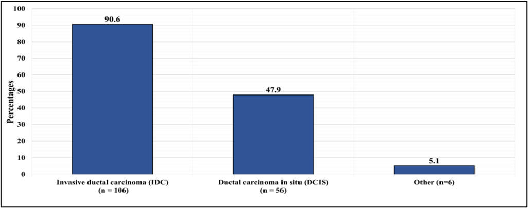
IDC was documented in 106 (90.6%), DCIS in 56 (42.7%.) The less common other histological subtypes accounted for 6 (5%). (Figure 1)
Reviewing the tumor characteristics in detail revealed: Grade I in 13 (11.1%), Grade II in 48 (41%), and Grade III in 52 (44.4%), and undocumented histological grade in 4 (3.4%), Surgical margins were positive in 24 (20.5%) and negative in 90 (76.9%), undocumented in 3(2.6%). All positive surgical margins underwent a second surgical procedure to ensure negativity. Lympho-vascular invasion (LVI) was present in 31(26.5%), absent in 74 (63.2%), and undocumented in 2 (10.3%). As for the nodal status, 56 (47.9%) were reported as node-negative, and 61 (52.1%) were reported as node-positive. The node positive group were further classified into with 1 positive lymph node (LN) in 14 (12%), 2 – 3 LNs in 20 (17.1%) and > 4 in 27 (23.1%).
On the immunohistochemical analysis reported ER was positive in 83 (70.9%), PR was positive in 66 (56.4%), and HER2 was positive in 35 (29.9%). TNBC was diagnosed in 17 (14.5%).
Ki67 level was performed in a limited number of the included group. 55 (47%) out of which the indices measurements were low index in 6 patients (5.1%), moderate in 15 (12.8%), and high in 34 (29.1%). Tumor size measurement by both imaging and pathology specimens have shown similar results. Lesions in the extreme of measurement <2 cm or >5 cm were reported with similar incidences of 14.5%, while lesions 2-5cm were reported in more than 50% of cases.
Table 2 Tumor Characteristics (n=117)
| n | % | |
|---|---|---|
| Histological grade | ||
| I | 13 | 11.1 |
| II | 48 | 41 |
| III | 52 | 44.4 |
| Missing | 4 | 3.4 |
| Surgical margins status | ||
| Positive | 24 | 20.5 |
| Negative | 90 | 76.9 |
| Missing | 3 | 2.6 |
| Lympho-vascular invasion | ||
| Present | 31 | 26.5 |
| Absent | 74 | 63.2 |
| Missing | 12 | 10.3 |
| Number of positive nodes | ||
| 0 | 56 | 47.90 |
| 1 | 14 | 12.00 |
| 2 - 3 | 20 | 17.10 |
| ≥ 4 | 27 | 23.10 |
| ER status | ||
| Positive | 83 | 70.9 |
| Negative | 34 | 29.1 |
| PR status | ||
| Positive | 66 | 56.4 |
| Negative | 50 | 42.7 |
| Missing | 1 | 0.9 |
| HER2 status | ||
| Positive | 35 | 29.9 |
| Negative | 81 | 69.2 |
| Missing | 1 | 0.9 |
| TNBC diagnosis | ||
| Yes | 17 | 14.5 |
| No | 100 | 85.4 |
| Ki67 | ||
| Low (<15%) | 6 | 5.1 |
| Moderate (15-30%) | 15 | 12.8 |
| High (>30%) | 34 | 29.1 |
| Missing | 62 | 53 |
| Radiological tumor size | ||
| <2 cm | 27 | 23.1 |
| 2 – 5 cm | 58 | 49.6 |
| >5 cm | 16 | 13.7 |
| Missing | 16 | 13.7 |
| Pathological tumor size | ||
| <2 cm | 17 | 14.5 |
| 2 – 5 cm | 66 | 56.4 |
| >5 cm | 17 | 14.5 |
| Missing | 17 | 14.5 |
67 patients (57.3%) underwent mastectomy, while 50 (42.7%) had undergone breast conserving surgery (BCS). Neoadjuvant therapy was administered in 38 (32.5%), while adjuvant therapy was given in the form of hormonal therapy in 93 (79.5%), radiotherapy in 74 (63.2%), and chemotherapy in 59 (50.4%) . (Table 3)
Table 3 Treatment Received (n = 117)
| n | % | |
|---|---|---|
| Neoadjuvant therapy | ||
| Yes | 38 | 32.5 |
| No | 78 | 66.7 |
| Missing | 1 | 0.9 |
| Surgical treatment | ||
| Mastectomy | 67 | 57.3 |
| BCS | 50 | 42.7 |
| Adjuvant radiotherapy | ||
| Yes | 74 | 63.2 |
| No | 41 | 35 |
| Missing | 2 | 1.7 |
| Adjuvant chemotherapy | ||
| Yes | 59 | 50.40 |
| No | 56 | 47.90 |
| Missing | 2 | 1.70 |
| Adjuvant hormonal therapy | ||
| Yes | 93 | 79.5 |
| No | 24 | 20.5 |
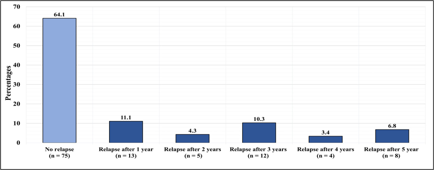
The variability in timing of relapse was also observed, with slight increase 13 (11.1%) in the first year compared to subsequent years; 5 (4.3%) in the second year, 12 (10.3%) in the third year, 4 (3.4%) in the fourth year, and 8 (6.8%) in the fifth year. (Figure-2)
The statistical univariate logistic regression was utilized in an attempt to delineate the pertinent factors that may predict relapse in BC. Variables that my contribute to disease relapse were explored, these included age, tumor size, tumor grade, margins status, the presences of LVI, nodal status and histological type.
Contrary to what was expected regarding the above listed pathological variables as potential cause for disease relapse, only age equal or less than 40 years (p = 0.04, odds ratio = 2.61), tumor size larger than 5 cm (T3) (p = 0.044, odds ratio = 4.4), , and TNBC (p = 0.039, odds ratio = 3.04) have shown higher tendency as risk factors for relapse. (Table 4)
Results from this study have failed in exhibiting the remaining variables as potential risk factors for relapse. This can be attributed to the small sample size and the inconsistency in reporting and documentation.
Table 4 Univariate Logistic Regression (Factors Predicting Relapse
| Factor | P value | OR | 95% CI |
|---|---|---|---|
| Age ≤40 years | 0.040* | 2.61 | 1.05 – 6.51 |
| Grade | |||
| II | 0.212 | 0.41 | 0.10 – 1.66 |
| III | 0.173 | 0.56 | 0.25 – 1.29 |
| Surgical margins status | 0.111 | 0.48 | 0.19 – 1.19 |
| LVI | 0.762 | 0.87 | 0.36 – 2.11 |
| Nodal status | |||
| 1 LN involved | 0.511 | 0.63 | 0.16 – 2.53 |
| 2 - 3 LN involved | 0.120 | 2.29 | 0.81 – 6.53 |
| 4 or more LN involved | 0.210 | 1.84 | 0.71 – 4.74 |
| ER status | 0.447 | 1.38 | 0.61 – 3.12 |
| PR status | 0.792 | 0.90 | 0.42 – 1.95 |
| HER2 status | 0.318 | 1.55 | 0.66 – 3.66 |
| TNBC | 0.039* | 3.04 | 1.06 – 8.70 |
| Ki67 (low is the referent) | |||
| Moderate | 0.149 | 0.14 | 0.01 – 2.01 |
| High | 0.624 | 1.58 | 0.25 – 9.82 |
| IDC | 0.534 | 0.64 | 0.16 – 2.57 |
| DCIS | 0.233 | 1.59 | 0.74 – 3.42 |
| Pathological tumor size, (< 2 cm is the referent) | |||
| (2 - 5 cm) | 0.758 | 1.20 | 0.38 – 3.84 |
| (>5 cm) | 0.044* | 4.40 | 1.04 – 18.60 |
| Surgical treatment (Mastectomy vs BCS) | 0.712 | 0.87 | 0.40 – 1.86 |
* Significant at level 0.05
4 Discussion#
Documented evidence in many validated studies have reported that the highest rates of disease relapse occur within the first 5 years after the initial treatment. Hence, a structured 5-year follow-up program was established and implemented. The rapid and promising evolution of advancing therapeutics and surgical options may have helped in the reduction of the disease relapse. Timely identification of patients with risk factors for relapse may aid in improving the clinical outcomes[10,23].
Age plays a role in the clinical presentation as younger women tend to present with larger and more aggressive tumors, rendering a higher risk of recurrence. Contrary to western, reports the patients in this review were younger, the majority were diagnosed in the fourth and fifth decades (61.5%), with a mean age at diagnosis of 49.6 years.
Reports have stated that concomitant DCIS maybe an independent prognostic factor for BC survival, with a positive impact in ER-positive patients.[24, 25] Other reports indicated that IDC and high-grade DCIS increases the chance of relapse by 2.5-folds with no effect on the overall survival.[26]
Confirming the results in the present study, with regard tumor size, reports have shown that larger lesion of > 5cm have higher tendency to relapse, rendering the tumor size as a sensible parameter for predicting recurrence.
Higher tumor grade has shown clinical significance as a strong prognostic indicator for enhancing the risk of recurrence.[27-29] However, other reports concluded that nuclear grade may not be an independent predictor of local invasive recurrence in DCIS.[30]
Data obtained in this current review have demonstrated high grade tumor in 100 (85.4%) yet, it failed to document its contribution to the statistical significance for relapse (p value=0.17).
Molecular classification of BC is based on IHC evaluation of receptor status has proven its significance in the choice of therapy, however, similar reports to the current study were unable to conclude its significant association with disease relapse, except in cases of TNBC[24, 32]
The timing of BC relapse in the in the first two and a half years maybe significantly correlated to Low levels of ER receptors[33].
Previous reports revealed no significant differences in overall survival (OS) and distant-disease free interval (DDFI) among patients who underwent mastectomy versus BCS in the first 18 months, however, more LRR was reported at 5 and 10 years in the BCS group.[33]
No statistically significant difference in relapse of BC (P=0.712) was documented in this study when comparing the type of the surgical procedure in the 5 years follow up.
Contrary to the results in the current review, multiple studies have demonstrated that a higher number of LN involvement (more than 3 LNs affected) was statistically correlated with a greater risk of LRR. Moreover, other studies have demonstrated that nodal involvement was independently associated with BC relapse.[34,35]
Microscopic positive surgical margins have always been the concern of surgeons, which has been documented as a strong risk factor for higher LRR risk and difference on OS and DDFI.[36] However, again this could not be demonstrated in this review(P=0.111).
Proposed additional indicators such as CRP (C-reactive protein) and SAA (serum amyloid A) ,CA15-3 , phosphatase (ALP), have demonstrated some potential in disclosing early recurrence.[37,38] T helper cells type 1 type 2 cytokines maybe also be able to reflect the BC immunosuppression potential to predict the probability of BC recurrence.[39]
It may appear that the recurrence rate in this review is higher than many published reports. This was expected considering the combined factors of unfavorable pathological characteristics present in each case. However, local recurrence occurred in only one third of patients 42(35.9%) while the remaining 75 (64.1%) did not relapse despite the similarity in characteristics
5 Limitations#
Many limitations were encountered in the process of data collection, namely, the small sample size, the inconsistency in investigations, radiological, and pathological reports, incomplete documentations and loss of follow up. In addition, the records were not designed for clinical studies.
6 Recommendations#
It is recommended that standardized patients ‘work up and management coupled with Accurate reporting and documentation, and establishing a wide database by encouraging multi-center studies with aid in accuracy of reports.
7 Conclusion#
BC relapse remains a challenging dilemma in the multidisciplinary treatment set up. The several pertinent prognostic BC indicators described in the literature individually or in combination have not accurately predicted disease outcome in every case, yet they have proven their relevance in defining various therapeutic modalities and treatment response.
Article information
- Published
Copyright © 2025 Maha Abdel Had. MD. This is an open access article distributed under the Creative Commons Attribution License, which permits unrestricted use, distribution, and reproduction in any medium, provided the original work is properly cited. CC BY 4.0 · Corrections policy
References 40#
- 1
Alqahtani W, Almufareh N, Domiaty D, Albasher G, Alduwish M, Alkhalaf H et al. Epidemiology of cancer in Saudi Arabia thru 2010–2019: a systematic review with constrained meta-analysis. AIMS Public Health. 2020;7(3):679-696.
- 2
Chaudhri E, Fathi W, Hussain F, Hashmi S. The Increasing Trends in Cases of the Most Common Cancers in Saudi Arabia. J Epidemiol Glob Health. 2020;10(4):258–262.
- 3
Saudi Cancer Registry Annual Report [Internet]. National Health Information Center. 2017 [cited 27 March 2022]. Available from: https://nhic.gov.sa/eServices/Documents/2017.pdf
- 4
Cardoso F, Kyriakides S, Ohno S, Penault-Llorca F, Poortmans P, Rubio I et al. Early breast cancer: ESMO Clinical Practice Guidelines for diagnosis, treatment and follow-up. Ann Oncol. 2019;30(8):1194-1220.
- 5
Babiker S, Nasir O, Alotaibi S, Marzogi A, Bogari M, Alghamdi T. Prospective breast cancer risk factors prediction in Saudi women. Saudi J Biol Sc. 2020;27(6):1624-1631
- 6
Hofvind S, Holen Å, Aas T, Roman M, Sebuødegård S, Akslen LA. Women treated with breast conserving surgery do better than those with mastectomy independent of detection mode, prognostic and predictive tumor characteristics. Eur J Surg Oncol. 2015 Oct;41(10):1417-22. doi: 10.1016/j.ejso.2015.07.002. Epub 2015 Jul 17. PMID: 26253193.
- 7
Wöckel A, Albert U, Janni W, Scharl A, Kreienberg R, Stüber T. The Screening, Diagnosis, Treatment, and Follow-Up of Breast Cancer. Dtsch Arztebl Int. 2018;115:316-23.
- 8
Ess S, Herrmann C, Bouchardy C, Neyroud I, Rapiti E, Konzelmann I et al. Impact of subtypes and comorbidities on breast cancer relapse and survival in population-based studies. Breast. 2018;41:151-158.
- 9
Viot J, Bachour M, Meurisse A, Pivot X, Fiteni F. Follow-up of patients with localized breast cancer and first indicators of advanced breast cancer recurrence: A retrospective study. Breast. 2017;34:53-57.
- 10
Ogawa Y, Ikeda K, Izumi T, Okuma S, Ichiki M, Ikeya T et al. First indicators of relapse in breast cancer: evaluation of the follow-up program at our hospital. Int J Clin Oncol. 2013;18(3):447-453.
- 11
Almagro E, González CS, Espinosa E. Factores pronósticos en el cáncer de mama en estadio inicial [Prognostic factors of early breast cancer]. Med Clin (Barc). 2016 Feb 19;146(4):167-71. Spanish. doi: 10.1016/j.medcli.2014.12.019. Epub 2015 Feb 26. PMID: 25726309.
- 12
Gulbahce HE, Downs-Kelly E, Herget KA, Stoddard GJ. The 21-Gene Recurrence Score in Special Histologic Subtypes of Breast Cancer. Arch Pathol Lab Med. 2022 Apr 1;146(4):478-484. doi: 10.5858/arpa.2020-0837-OA. PMID: 34343231.
- 13
Schaafsma E, Zhang B, Schaafsma M, Tong CY, Zhang L, Cheng C. Impact of Oncotype DX testing on ER+ breast cancer treatment and survival in the first decade of use. Breast Cancer Res. 2021 Jul 17;23(1):74. doi: 10.1186/s13058-021-01453-4. PMID: 34274003; PMCID: PMC8285794.
- 14
Mao N, Yin P, Zhang H, Zhang K, Song X, Xing D, Chu T. Mammography-based radiomics for predicting the risk of breast cancer recurrence: a multicenter study. Br J Radiol. 2021 Nov 1;94(1127):20210348. doi: 10.1259/bjr.20210348. Epub 2021 Sep 14. PMID: 34520235; PMCID: PMC8553203.
- 15
Pan H, Gray R, Braybrooke J, Davies C, Taylor C, McGale P, Peto R, Pritchard KI, Bergh J, Dowsett M, Hayes DF; EBCTCG. 20-Year Risks of Breast-Cancer Recurrence after Stopping Endocrine Therapy at 5 Years. N Engl J Med. 2017 Nov 9;377(19):1836-1846. doi: 10.1056/NEJMoa1701830. PMID: 29117498; PMCID: PMC5734609.
- 16
Wapnir IL, Khan A. Current Strategies for the Management of Locoregional Breast Cancer Recurrence. Oncology (Williston Park). 2019 Jan 17;33(1):19-25. PMID: 30731014.
- 17
Chen H, Bai F, Wang M, Zhang M, Zhang P, Wu K. The prognostic significance of co-existence ductal carcinoma in situ in invasive ductal breast cancer: a large population-based study and a matched case-control analysis. Ann Transl Med. 2019;7(18):484.
- 18
10.3389/fonc.2021.734719. PMID: 34888233; PMCID: PMC8650120
- 19
Elkum N, Dermime S, Ajarim D, Al-Zahrani A, Alsayed A, Tulbah A et al. Being 40 or younger is an independent risk factor for relapse in operable breast cancer patients: The Saudi Arabia experience. BMC Cancer. 2007;7:222.
- 20
Mo C, Ruan W, Lin J, Chen H, Chen X. Repeat Breast-Conserving Surgery Versus Salvage Mastectomy for Ipsilateral Breast Tumour Recurrence After Breast-Conserving Surgery in Breast Cancer Patients: A Meta-Analysis. Front Oncol. 2021 Nov 23;11:734719. doi: 10.3389/fonc.2021.734719. PMID: 34888233; PMCID: PMC8650120
- 21
Han W, Kang S. Relationship between age at diagnosis and outcome of premenopausal breast cancer: age less than 35 years is a reasonable cut-off for defining young age-onset breast cancer. Breast Cancer Res Treat. 2010;119(1):193-200.
- 22
Fowble B, Schultz D, Overmoyer B, Solin L, Fox K, Jardines L et al. The influence of young age on out- come in early stage breast cancer. Int J Radiat Oncol Biol Phys. 1994;30:23-33.
- 23
Martínez M, Oltra S, Peña-Chilet M, Alonso E, Hernando C, Burgues O et al. Breast Cancer in Very Young Patients in a Spanish Cohort: Age as an Independent Bad Prognostic Indicator. Breast Cancer (Auckl). 2019;13:297–305.
- 24
Arvold N, Taghian A, Niemierko A, Abi Raad R, Sreedhara M, Nguyen P. Age, Breast Cancer Subtype Approximation, and Local Recurrence After Breast-Conserving Therapy. J Clin Oncol. 2011;29:3885–91.
- 25
Braunstein L, Taghian A, Niemierko A, Salama L, Capuco A, Bellon J et al. Breast-cancer subtype, age, and lymph node status as predictors of local recurrence following breast-conserving therapy. Breast Cancer Res Treat. 2016;161(1):173-179.
- 26
Chen H, Bai F, Wang M, Zhang M, Zhang P, Wu K. The prognostic significance of co-existence ductal carcinoma in situ in invasive ductal breast cancer: a large population-based study and a matched case-control analysis. Ann Transl Med. 2019;7(18):484.
- 27
Kim J, Han W, Moon H, Park I, Ahn S, Kim J et al. Grade of Ductal Carcinoma In Situ Accompanying Infiltrating Ductal Carcinoma As an Independent Prognostic Factor. Clin Breast Cancer. 2013;13(5):385-391
- 28
Wang X, Zhang L, Zhang X, Luo J, Wang X, Chen X et al. Impact of clinical-pathological factors on locoregional recurrence in mastectomy patients with T1-2N1 breast cancer: who can omit adjuvant radiotherapy?. Breast Cancer Res Treat. 2021;190(2):277-286.
- 29
Asaoka M, Narui K, Suganuma N, Chishima T, Yamada A, Sugae S et al. Clinical and pathological predictors of recurrence in breast cancer patients achieving pathological complete response to neoadjuvant chemotherapy. Eur J Surg Oncol. 2019;45(12):2289-2294.
- 30
Maishman T, Cutress R, Hernandez A, Gerty S, Copson E, Durcan L et al. Local Recurrence and Breast Oncological Surgery in Young Women With Breast Cancer. Ann Surg. 2017;266(1):165-172.
- 31
Kim J, Park K, Kang G, Kim H, Gwak G, Shin Y. Predictors of Recurrent Ductal Carcinoma In Situ after Breast-Conserving Surgery. J Breast Cancer. 2016;19(2):185.
- 32
Zhang X, Dai H, Liu B, Song F, Chen K. Predictors for local invasive recurrence of ductal carcinoma in situ of the breast. Eur J Cancer Prev. 2016;25(1):19-28.
- 33
Falco M, Masojć B, Kram A. Locoregional relapse is a strong prognostic indicator of distant metastatic progression in breast cancer patients after negative sentinel lymph node biopsy. Breast J. 2020;26(12):2364-2370.
- 34
Cao jJ, Olson R, Tyldesley S. Comparison of Recurrence and Survival Rates after Breast-Conserving Therapy and Mastectomy in Young Women with Breast Cancer. Curr Oncol. 2013;20(6):593-601.
- 35
Kennecke H, McArthur H, Olivotto I, Speers C, Bajdik C, Chia S et al. Risk of early recurrence among postmenopausal women with estrogen receptor-positive early breast cancer treated with adjuvant tamoxifen. Cancer. 2008;112(7):1437-1444.
- 36
Cho H, Van Belle V, Vandorpe T, Wildiers H, Janssen H, Leunen K et al. Prognostic Significance of Nodal and PgR Status on Early Relapse in Operable HER-2 Positive Breast Cancer from the Pre-Trastuzumab Era. Cancer Res. 2009;69(24):6046.
- 37
Houssami N, Macaskill P, Luke Marinovich M, Morrow M. The Association of Surgical Margins and Local Recurrence in Women with Early-Stage Invasive Breast Cancer Treated with Breast-Conserving Therapy: A Meta-Analysis. Ann Surg Oncol. 2014;21(3):717-730.
- 38
Keshaviah A, Dellapasqua S, Rotmensz N, Lindtner J, Crivellari D, Collins J, et al. CA15-3 and alkaline phosphatase as predictors for breast cancer recurrence: A combined analysis of seven International Breast Cancer Study Group trials. Ann Oncol. 2007;18(4).
- 39
McAndrew N, Bottalico L, Mesaros C, Blair I, Tsao P, Rosado J, et al. Effects of systemic inflammation on relapse in early breast cancer. npj Breast Cancer. 2021;7(1).
- 40
Wang L, Simons D, Lu X, Tu T, Solomon S, Wang R et al. Connecting blood and intratumoral Treg cell activity in predicting future relapse in breast cancer. Nat Immunol. 2019;20(9):1220-1230.