Seroprevalence Of Helicobacter pylori Among Patients Attending Ignatius Ajuru University Of Education Medical Centre
Uzah G. A.
Uzah G. A. More articles by this author →Chuks Dike B.
Chuks Dike B. More articles by this author →Nwaokeleme E. B.
Nwaokeleme E. B. More articles by this author →
Published
Abstract
Helicobacter pylori infection remains a major global health concern, particularly in developing countries where poor sanitation and overcrowding contribute to high prevalence levels. This study determined the seroprevalence of H. pylori infection among patients attending the Ignatius Ajuru University of Education (IAUE) Medical Centre, Rivers State, Nigeria, and examined associated sociodemographic and behavioural risk factors. A cross-sectional descriptive design was employed, involving 98 consenting patients from whom blood samples were collected and analyzed using enzyme-linked immunosorbent assay (ELISA) to detect H. pylori-specific IgG antibodies. Relevant sociodemographic and risk-related data were obtained through structured questionnaires and analyzed using descriptive and inferential statistics. Findings revealed an overall seroprevalence of 57.1%, indicating a high burden of H. pylori infection within the study population. Age showed a statistically significant association with infection (p = 0.048), with individuals aged 26–35 years having the highest prevalence. Sex was not significantly associated with H. pylori seropositivity (p = 0.73). Among assessed behavioural factors, sharing of eating utensils demonstrated a significant association with H. pylori infection (p = 0.026), while handwashing before meals showed no significant association (p = 0.09). These results highlight the role of close interpersonal contact in H. pylori transmission. The study concludes that H. pylori infection is highly prevalent among patients attending IAUE Medical Centre and that age and sharing of eating utensils are significant predictors of infection. It recommends awareness campaigns, routine screening, behavioural modification to discourage utensil sharing, and improved access to diagnostic services. As the first documented assessment of H. pylori burden in this setting, the study provides baseline data that will guide future research and inform public health interventions aimed at reducing associated gastrointestinal complications.
1 Introduction#
Helicobacter pylori (H. pylori) is a Gram-negative, microaerophilic bacterium found in the stomach, strongly associated with gastritis, peptic ulcers, and gastric cancer. It infects more than half of the world’s population, with a higher prevalence in developing countries due to factors like poor sanitation and overcrowding (Hooi et al., 2017; Zamani et al., 2018). Recent data suggest that regional variations in H. pylori prevalence remain substantial, with sub-Saharan Africa reporting some of the highest infection rates globally (Eusebi et al., 2020).
Helicobacter pylori (H. pylori) is one of the most prevalent chronic bacterial infections worldwide. This spiral-shaped, Gram-negative bacterium predominantly colonizes the gastric mucosa and has been implicated in the pathogenesis of several upper gastrointestinal diseases such as chronic gastritis, duodenal and gastric ulcers, mucosa-associated lymphoid tissue (MALT) lymphoma, and gastric cancer (Malfertheiner et al., 2023; Eusebi et al., 2014). The organism has a remarkable ability to survive the acidic environment of the stomach, primarily through urease production, which neutralizes gastric acid and facilitates colonization.
Globally, billions of individuals are estimated to be infected with H. pylori, with marked regional differences in prevalence. The highest prevalence rates are reported in Africa, Latin America, and parts of Asia, where infection rates often exceed 70% (Hooi et al., 2017; Zamani et al., 2018). These disparities are closely linked to socioeconomic conditions, sanitation, access to clean water, and crowding, which are conducive to the bacterium fecal–oral and oral–oral transmission pathways (Vale & Vítor, 2010; Duan et al. 2023; Ekundayo et al. 2023).
In Nigeria, H. pylori infection remains a significant public health issue, with multiple hospital-based studies showing high prevalence among symptomatic and asymptomatic patients. Recent findings have reported seroprevalence ranging between 30% and 85% among various population groups in Nigeria and Africa (Lawson, 2023; Palamides et al., 2020; Ray-Offor & Obiorah, 2018; Smith et al., 2022). Despite this burden, routine testing is not common in many primary care settings due to cost and infrastructure limitations, leaving many cases undiagnosed or misdiagnosed.
Within the Rivers State region, there is a growing concern about the underreporting and underdiagnosis of H. pylori infections. A few studies in Port Harcourt have documented high infection rates among patients with dyspeptic symptoms, highlighting the need for localized surveillance and intervention (Lawson, 2023). However, there is a paucity of data on infection rates specifically within tertiary institutions and their healthcare centers, including the IAUE Medical Centre, which caters to a large number of students and staff.
Students and staff in university environments like IAUE may face unique risk factors such as shared accommodation, poor dietary habits, and inadequate hygiene practices, which could predispose them to H. pylori infection (Vale & Vítor, 2010; Duan et al. 2023; Ekundayo et al. 2023). Yet, no study to date has systematically evaluated the seroprevalence of H. pylori within this academic medical setting. Establishing accurate prevalence data is critical for informing health promotion strategies, diagnostic protocols, and appropriate therapeutic interventions.
Moreover, the emergence of antibiotic-resistant strains of H. pylori further underscores the importance of surveillance. Treatment failure due to increasing antimicrobial resistance, including resistance to clarithromycin and metronidazole, represents a major challenge in H. pylori eradication, posing a challenge to effective eradication (Harrison et al., 2017; Savoldi et al., 2018; Chukwudike et al., 2024). Understanding the seroprevalence or infection rates and potential risk factors in specific populations such as that served by IAUE Medical Centre can contribute to the global understanding of H. pylori epidemiology and improve patient outcomes through tailored public health responses and improving clinical management strategies.
2 Methodology#
2.1 Study Design#
A cross-sectional descriptive design was adopted in this study to determine the seroprevalence of Helicobacter pylori infection among patients attending the medical outpatient clinic at Ignatius Ajuru University of Education (IAUE) Medical Centre, Rivers State, Nigeria. The cross-sectional approach is appropriate for estimating the prevalence of a condition at a single point in time and for exploring associations between H. pylori infection and various sociodemographic or clinical variables within the target population.
2.2 Study Area#
The study was conducted at the Medical Centre of Ignatius Ajuru University of Education (IAUE), located in Rumuolumeni, Port Harcourt, Rivers State, Nigeria. IAUE is a public university established to provide higher education and promote research across various academic disciplines. The university operates a medical centre that offers primary and secondary healthcare services to its students, staff, and members of the surrounding communities. The IAUE Medical Centre is equipped with basic diagnostic facilities, a pharmacy, consulting rooms, and a laboratory that supports routine clinical investigations.
Port Harcourt, the capital of Rivers State, is a major urban center in southern Nigeria with a diverse population and a mix of urban and peri-urban settlements. Rivers State lies in the Niger Delta region, which is characterized by rapid urbanization, oil-based industrial activity, and a tropical climate. The state faces various public health challenges, including limited access to clean water and sanitation, both of which are important risk factors for Helicobacter pylori transmission. These environmental and socioeconomic conditions make the area particularly suitable for investigating the burden of H. pylori infection.
The IAUE Medical Centre was selected for this study due to its accessibility to a broad patient population that includes individuals from both academic and non-academic backgrounds. The Centre receives patients with varied medical complaints, including gastrointestinal symptoms, making it an appropriate setting for assessing the seroprevalence of H. pylori. Additionally, the university environment presents an opportunity to evaluate the infection in a relatively defined and structured population, which can contribute valuable insights for targeted health interventions and future research.
2.3 Study Population#
The study population consists of consenting adult patients (aged ≥18 years) attending the IAUE Medical Centre for general medical consultation during the study period. Patients presenting with gastrointestinal symptoms suggestive of dyspepsia (e.g., epigastric pain, bloating, nausea) were prioritized, although asymptomatic individuals were also included to capture broader community-level infection rates.
2.4 Inclusion Criteria#
- Patients aged ≥18 years.
- Willing to provide informed consent.
2.5 Exclusion Criteria#
Patients on recent antibiotic or proton pump inhibitor therapy (within the past 2 weeks), previous H. pylori treatment, or chronic comorbidities that could confound test results (e.g., malignancies or immunosuppressive conditions) were excluded from the study.
2.6 Sample Size Determination#
A total of 98 samples were collected from consenting patients attending IAUE Medical Centre. The sample size determination was based on standard prevalence estimation formulas, using an assumed H. pylori prevalence of 55% (based on prior regional studies), a 95% confidence interval, and a 5% margin of error.
Using the formula:
n=(Z2 pq)/d2
Where:
- Z = 1.96 (95% confidence)
- p = assumed prevalence (55%)
- q = 1 - p
- d = margin of error (0.05)
2.7 Sampling Technique#
Systematic random sampling of patients who meet the inclusion criteria were considered. Blood samples were collected under sterile conditions, and serum were tested using a commercially validated enzyme-linked immunosorbent assay (ELISA) to detect H. pylori-specific IgG antibodies. This serological method is cost-effective and suitable for epidemiological surveys, although it detects both current and past infections.
2.8 Data Collection and Analysis#
Data on sociodemographic characteristics, clinical symptoms, hygiene practices, and dietary habits were collected via structured questionnaires and analyzed using descriptive and inferential statistics (e.g., chi-square tests and logistic regression). A p-value <0.05 were considered statistically significant. The outcomes of the serological testing and sociodemographic data were corroborated to other research work and which help to inform local disease burden and guide public health interventions targeting H. pylori control.
2.9 Ethical Considerations#
Ethical approval was obtained from the IAUE Research Ethics Committee. Informed consent was secured from each participant, and confidentiality was maintained.
3 Results#
3.1 A.Sociodemographic characteristic of respondents#
Table 1.1 Age Distribution of Respondents.
| Age Group | Frequency (n) | Percentage (%) |
|---|---|---|
| 10 – 20 | 56 | 57.1 |
| 21 – 30 | 31 | 31.6 |
| 31 – 40 | 11 | 11.2 |
| Total | 98 | 100 |
Source: An Extract From SPSS Version 25.0
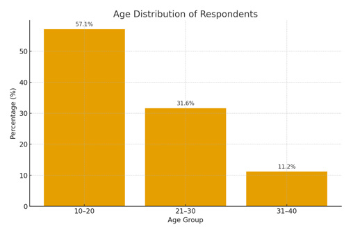
Table 1.1 and figure 1.1 shows the age distribution of the 98 respondents. The largest proportion (57.1%) falls within the 10–20 years age group, followed by 21–30 years with 31.6%. Only 11.2% of the respondents are between 31–40 years. This indicates that most participants were young individuals, likely reflecting the IAUE demographic dominated by students and young adults.
Table 1.2 Sex Distribution of Respondents.
| Sex | Frequency (n) | Percentage (%) |
|---|---|---|
| Male | 82 | 83.7 |
| Female | 16 | 16.3 |
| Total | 98 | 100 |
Source: An Extract From SPSS Version 25.0.
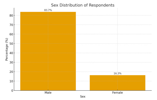
Table 1.2 and figure 1.2 shows the sex distribution of the 98 respondents. The majority of the respondents were male, accounting for 83.7% (n = 82), while females constituted only 16.3% (n = 16). This indicates that the study population was predominantly male, which may reflect the demographic characteristics of the IAUE Medical Centre attendees.
Table 1.3 Marital Status of Respondents.
| Marital Status | Frequency (n) | Percentage (%) |
|---|---|---|
| Single | 93 | 94.9 |
| Married | 5 | 5.1 |
| Divorced | 0 | 0.0 |
| Widowed | 0 | 0.0 |
| Total | 98 | 100 |
Source: An Extract From SPSS Version 25.0
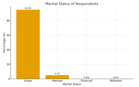
Table 1.3 and figure 1.3 shows the marital status distribution of the 98 respondents. The majority were single, accounting for 94.9% (n = 93), while only 5.1% (n = 5) were married. None of the respondents were divorced or widowed. This shows that most participants were young adults, which aligns with the IAUE Medical Centre population that mainly consists of students.
Table 1.4 Educational Level of Respondents.
| Educational Level | Frequency (n) | Percentage (%) |
|---|---|---|
| No Formal Education | 0 | 0.0 |
| Primary | 0 | 0.0 |
| Secondary | 20 | 20.4 |
| Tertiary | 78 | 79.6 |
| Total | 98 | 100 |
Source: An Extract From SPSS Version 25.0
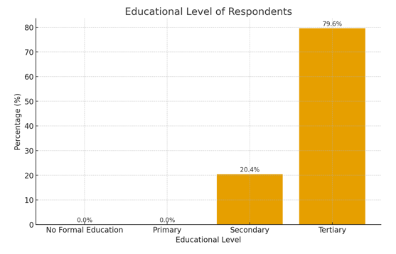
Table 1.4 and Figure 1.4 present the educational level of the 98 respondents. A majority (79.6%) had tertiary education, while 20.4% had secondary education. None of the respondents reported having primary or no formal education. This indicates that the study participants were predominantly well-educated, consistent with the IAUE Medical Centre population, which largely comprises students and staff of an academic institution.
Table 1.5 Occupation of Respondents.
| Occupation | Frequency (n) | Percentage (%) |
|---|---|---|
| Student | 90 | 91.8 |
| Others | 8 | 8.2 |
| Total | 98 | 100 |
Source: An Extract From SPSS Version 25.0
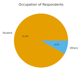
Table 1.5 and figure 1.5 show that the vast majority of respondents (91.8%) were students, while only 8.2% fell into the ‘Others’ category. This indicates that most participants were members of the IAUE academic environment, which aligns with the study setting at the IAUE Medical Centre. The dominance of students in the sample implies that the findings primarily reflect the health profile of a young, school-based population.
Table 1.6 Place of Residence of Respondents.
| Residence | Frequency (n) | Percentage (%) |
|---|---|---|
| Urban | 54 | 55.1 |
| Semi-urban | 30 | 30.6 |
| Rural | 14 | 14.3 |
| Total | 98 | 100 |
Source: An Extract From SPSS Version 25.0
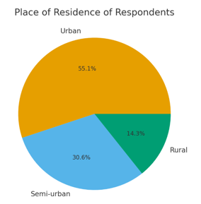
Table 1.6 and Figure 1.6 present the place of residence of the 98 respondents. A majority (55.1%) resided in urban areas, followed by 30.6% living in semi-urban locations. Only 14.3% were from rural areas. This distribution indicates that most participants were urban dwellers, which aligns with the IAUE Medical Centre’s location within an urban environment. The predominance of urban residents suggests easier access to healthcare services, which may influence the study’s findings.
Table 1.7 Monthly Household Income of Respondents
| Monthly Income ₦ | Frequency (n) | Percentage (%) |
|---|---|---|
| < 50,000 | 48 | 49.0 |
| 50,000 – 100,000 | 21 | 21.4 |
| 100,001 – 200,000 | 14 | 14.3 |
| > 200,000 | 15 | 15.3 |
| Total | 98 | 100 |
Source: An Extract From SPSS Version 25.0
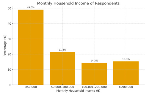
Table 1.7 and Figure 1.7 present the monthly household income of the 98 respondents. Nearly half (49.0%) reported earning less than ₦50,000 monthly, while 21.4% earned between ₦50,000 and ₦100,000. Additionally, 14.3% earned between ₦100,001 and ₦200,000, and 15.3% reported earnings above ₦200,000. This distribution shows that most respondents fall within the lower-income category, which may reflect the socioeconomic characteristics of the IAUE Medical Centre population, comprising mainly students and lower-income earners.
3.2 B.Seroprevalence of H. pylori Infection#
Table 2.1 H. pylori Seropositivity among Patients.
| H. pylori Status | Frequency | Percentage (%) |
|---|---|---|
| Positive | 56 | 57.1 |
| Negative | 42 | 42.9 |
| Total | 98 | 100.0 |
Seroprevalence = 57.1%
Table 2.1 shows that out of the 98 patients tested for Helicobacter pylori infection, 56 (57.1%) were seropositive, while 42 (42.9%) tested negative. This indicates that more than half of the sampled population had been exposed to or infected with H. pylori. The seroprevalence rate of 57.1% suggests that H. pylori infection is relatively common among patients attending the IAUE Medical Centre and may represent a significant public health concern requiring further attention in terms of prevention, diagnosis, and treatment strategies.
Table 2.2 Age vs H. pylori Seropositivity.
| Age Group | Positive | Negative | Total |
|---|---|---|---|
| 18–25 | 15 | 16 | 31 |
| 26–35 | 22 | 15 | 37 |
| 36–45 | 10 | 7 | 17 |
| 46–55 | 5 | 3 | 8 |
| >55 | 4 | 1 | 5 |
| Total | 56 | 42 | 98 |
χ2 = 6.03, p = 0.048 (significant)
Table 2.2 presents the relationship between age and H. pylori seropositivity among the 98 respondents. The highest number of positive cases occurred in the 26–35 age group (22 out of 37), followed by the 18–25 group (15 out of 31). The lowest frequency of positive cases was found among respondents above 55 years (4 out of 5). The chi-square test result (χ2 = 6.03, p = 0.048) indicates that there is a statistically significant association between age and H. pylori infection at the 0.05 level. This suggests that H. pylori seropositivity varies significantly across age categories, with younger adults especially those aged 26–35 appearing more affected than older participants.
Table 2.3 Sex vs H. pylori Seropositivity.
| Sex | Positive | Negative | Total |
|---|---|---|---|
| Male | 25 | 20 | 45 |
| Female | 31 | 22 | 53 |
| Total | 56 | 42 | 98 |
χ2 = 0.12, p = 0.73 (not significant)
Table 2.3 shows the relationship between sex and H. pylori seropositivity. Among the 45 males, 25 tested positives, while 31 out of 53 females were positive for H. pylori. Although more females were infected than males in absolute numbers, the chi-square test result (χ2 = 0.12, p = 0.73) indicates no statistically significant association between sex and H. pylori infection. This means that infection rates did not differ meaningfully by sex, suggesting that both males and females have a comparable likelihood of acquiring H. pylori within the study population.
Table 2.4 Hygiene Practices vs H. pylori Infection
| Handwashing Before Meals | Positive | Negative | Total |
|---|---|---|---|
| Yes | 32 | 28 | 60 |
| No | 24 | 14 | 38 |
| Total | 56 | 42 | 98 |
χ2 = 2.18, p = 0.09 (not significant)
Table 2.4 examines the relationship between handwashing practices before meals and H. pylori infection. Among respondents who regularly washed their hands before meals, 32 out of 60 tested positives, while 24 out of 38 who did not wash hands tested positive. Although a higher proportion of non-handwashers were infected, the chi-square result (χ2 = 2.18, p = 0.09) shows no statistically significant association between handwashing habits and H. pylori seropositivity at the 0.05 level. This suggests that while poor hygiene may contribute to infection risk, it was not a strong predictor of H. pylori infection in this study population.
Table 2.5 Sharing Eating Utensils vs H. pylori Infection.
| Sharing Eating Utensils | Positive | Negative | Total |
|---|---|---|---|
| Yes | 37 | 17 | 54 |
| No | 19 | 25 | 44 |
| Total | 56 | 42 | 98 |
χ2 = 4.95, p = 0.026 (significant)
Table 2.5 shows the relationship between sharing eating utensils and H. pylori infection. Among respondents who shared utensils, 37 out of 54 tested positive, compared to 19 out of 44 who did not share utensils. The chi-square test (χ2 = 4.95, p = 0.026) indicates a statistically significant association between sharing eating utensils and H. pylori seropositivity. This suggests that individuals who share eating utensils are more likely to be infected with H. pylori, highlighting shared utensils as a potential transmission route within the study population.
4 Discussion#
The study revealed a relatively high seroprevalence of Helicobacter pylori among patients attending IAUE Medical Centre, with 57.1% (56/98) testing positive for H. pylori antibodies. This finding indicates that more than half of the study population had previous exposure to or infection with the organism. The observed prevalence may be associated with environmental conditions, sanitation challenges, and socioeconomic factors that influence H. pylori transmission. Studies have shown that infection remains highly prevalent in developing regions, particularly in Africa, where childhood exposure, household transmission, and poor living conditions contribute to sustained infection rates with prevalence rates exceeding 70% (Hooi et al., 2017; Smith et al., 2022). Similarly, Nigerian studies have reported substantial levels of H. pylori infection among different populations, highlighting the continued public health importance of the organism in the country (Harrison et al., 2017; Palamides et al., 2020). The present finding suggests that H. pylori remains an important health concern in Rivers State.
However, this result is in contrast with the submission of Adim et al (2024) who reported a prevalence of 31% among pregnant women in Mbodo-Aluu community, indicating that H. pylori prevalence may vary with population characteristics. This finding is supported by epidemiological reviews have emphasized that sanitation, household crowding, water contamination, and socioeconomic conditions contribute significantly to differences in H. pylori prevalence worldwide (Mežmale et al., 2020; Zamani et al., 2018).
Furthermore, the study revealed a significant relationship between age and H. pylori infection (p = 0.048). Respondents aged 26–35 years accounted for the highest number of positive cases, followed by those aged 18–25 years. This suggests increased exposure among young adults, which may be related to lifestyle patterns, social interaction possibly due to higher movement, shared accommodation, poor dietary habits, and work-related stress. Previous studies indicate that H. pylori acquisition commonly occurs early in life and infection patterns may reflect cumulative exposure to risk factors throughout life (Eusebi et al., 2014; Zamani et al., 2018). However, the present finding differs from reports suggesting increasing prevalence with advancing age, supported by the life-course epidemiology perspective, which implies that exposures in early adulthood increase the likelihood of infection before immunity declines where older individuals demonstrate higher infection rates due to longer duration of exposure (Hooi et al., 2017; Eusebi et al., 2014).
However, the study revealed no significant relationship between sex and H. pylori seropositivity (p = 0.73). Although females (31) had slightly higher infection rates than males (25), the difference was not statistically significant. This suggests that sex does not play a meaningful role in determining H. pylori susceptibility among patients attending IAUE Medical Centre. This finding is consistent with Hooi et al. (2017) who reported that H. pylori infection shows no sex predilection. Similarly, Smith et al. (2018), which investigated clinical and sociodemographic risk factors for H. pylori infection in Nigeria and found no significant association between sex and infection status. The study reported similar infection proportions between males and females. The present result reflects the biological theory that exposure rather than biological sex is the major determinant of infection.
Similarly, no significant relationship was observed between handwashing before meals and H. pylori infection (p = 0.09) in the study. Although individuals with poor handwashing practices showed a higher proportion of infection, the association was not statistically significant. This finding supports the understanding that H. pylori transmission is complex and involves multiple pathways, including oral–oral, fecal–oral, and environmental routes (Duan et al., 2023; Vale & Vítor, 2010). Previous studies have indicated that hygiene practices may influence transmission; however, handwashing alone may not completely prevent infection because exposure may occur through contaminated food, water, and close household contact (Salih, 2009; Ekundayo et al., 2023). The finding is supported by public health theory which proposes that disease transmission is multifactorial and cannot be attributed to a single behavioral factor.
Finally, the study revealed a significant relationship between sharing of eating utensils and H. pylori infection (p = 0.026). Participants who shared utensils recorded more positive cases than those who did not, suggesting that close interpersonal contact may contribute to transmission. This finding agrees with evidence that intrafamilial spread and oral–oral transmission pathways contribute to H. pylori dissemination (Vale & Vítor, 2010; Duan et al., 2023). The finding also supports previous observations that household behaviors and communal practices may influence infection risk. However, transmission is multifactorial, and environmental factors such as sanitation and water quality may also contribute substantially to infection patterns (Ekundayo et al., 2023; Mežmale et al., 2020). The present finding is supported by the infectious disease transmission model, which identifies direct contact as a significant route of transmission.
5 Conclusions#
The study established that H. pylori infection is highly prevalent among patients attending IAUE Medical Centre. The significant association between age and infection suggests that young adults may be at greater risk due to lifestyle characteristics such as communal living and poor hygiene practices. Although infection rates were slightly higher among females, sex showed no significant association with infection.
Importantly, sharing eating utensils was significantly associated with H. pylori seropositivity, indicating that oral–oral transmission remains a critical pathway within the study population. The lack of significant association with handwashing reinforces the multifactorial nature of H. pylori transmission, which may involve close contact, poor sanitation, and dietary habits.
Overall, the findings emphasize the need for improved awareness, early diagnosis, and behavioural modifications to reduce the burden of H. pylori infection in the IAUE community.
5.1 Disclaimer (Artificial Intelligence)#
Author(s) hereby declare that NO generative AI technologies such as Large Language Models (ChatGPT, COPILOT, etc.) and text-to-image generators have been used during the writing or editing of this manuscript.
Article information
- Published
Copyright © 2026 Gift Atumatuchukwu Uzah. This is an open access article distributed under the Creative Commons Attribution License, which permits unrestricted use, distribution, and reproduction in any medium, provided the original work is properly cited. CC BY 4.0 · Corrections policy
References 37#
- 1
Abdulrasheed, A., Lawal, O. O., Abioye-Kuteyi, E. A., & Lamikanra, A. (2005). Antimicrobial susceptibility of Helicobacter pylori isolates of dyspeptic Nigerian patients. Tropical Gastroenterology, 26(2), 85–88.
- 2
Adim, C.C., Nnamdi, F.H., Ugboma, C.D., et al. (2024). Detection and Prevalence of Syphilis, Hepatitis C Virus and Helicobacter pylori Co-Infection among Pregnant Women attending Primary Health Centre, Mbodo-Aluu, Rivers State, Nigeria. medRxiv. https://doi.org/10.1101/2024.03.22.24304749
- 3
Best, L. M. J., Takwoingi, Y., Siddique, S., Selladurai, A., Gandhi, A., Low, B., Yousaf, S., & Gurusamy, K. S. (2018). Non-invasive diagnostic tests for Helicobacter pylori infection. Cochrane Database of Systematic Reviews, 2018(3), CD012080. https://doi.org/10.1002/14651858.CD012080.pub2
- 4
Chukwudike, E.S., Asombang, A. W., Monteiro, J.F.G., Sawyer, K., Argueta, E., Tazinkeng, N .N. & Moss, S. F. (2024). Helicobacter pylori eradication rates and antibiotic resistance patterns in Nigeria: A systematic review and meta-analysis. American Journal of Gastroenterology and Hepatology, 5(1):1025.
- 5
Duan, M., Li, Y., Liu, J., Zhang, W., Dong, Y., Han, Z., Wan, M., Lin, M., Lin, B., Kong, Q., Ding, Y., Yang, X., Zuo, X., & Li, Y. (2023). Transmission routes and patterns of Helicobacter pylori. Helicobacter, 28(1), e12945. https://doi.org/10.1111/hel.12945.
- 6
Ekundayo, T. C., Swalaha, F. M. & Ijabadeniyi, O. A. (2023). Global and regional prevalence of Helicobacter pylori in drinking waters: A sustainable, human development and socio-demographic indices based meta-regression-modelling. (2023). Science of the Total Environment, 861, 160633. https://doi.org/10.1016/j.scitotenv.2022.160633.
- 7
Etukudo, O. M., Ikpeme, E. E., & Ekanem, E. E. (2012). Seroepidemiology of Helicobacter pylori infection among children seen in a tertiary hospital in Uyo, southern Nigeria. Pan African Medical Journal, 12, Article 39. https://doi.org/10.11604/pamj.2012.12.39.1377
- 8
Eusebi, L. H., Zagari, R. M., & Bazzoli, F. (2014). Epidemiology of Helicobacter pylori infection. Helicobacter, 19(Suppl. 1), 1–5. https://doi.org/10.1111/hel.12165
- 9
Fallone, C. A., Chiba, N., Veldhuyzen van Zanten, S., Fischbach, L., Gisbert, J. P., Hunt, R. H., Jones, N. L., Render, C., Leontiadis, G. I., Moayyedi, P., & Marshall, J. K. (2016). The Toronto Consensus for the Treatment of Helicobacter pylori Infection in Adults. Gastroenterology, 151(1), 51–69.e14. https://doi.org/10.1053/j.gastro.2016.04.006
- 10
Gisbert, J. P., & Calvet, X. (2019). Review: Diagnosis of Helicobacter pylori infection. Helicobacter, 24(Suppl. 1), Article e12641. https://doi.org/10.1111/hel.12641
- 11
Gottrup, F., Apelqvist, J., Bjarnsholt, T., Cooper, R., Moore, Z., Peters, E. J. G., & Probst, S. (2014). Antimicrobials and non-healing wounds. Evidence, controversies and suggestions—key messages. Journal of Wound Care, 23(10), 477–482. https://doi.org/10.12968/jowc.2014.23.10.477
- 12
Haque, R., Huston, C. D., Hughes, M., Houpt, E., & Petri, W. A., Jr. (2003). Amebiasis. The New England Journal of Medicine, 348(16), 1565–1573. https://doi.org/10.1056/NEJMra022710
- 13
Harrison, U., Fowora, M. A., Seriki, A. T., et al. (2017). Helicobacter pylori strains from a Nigerian cohort show divergent antibiotic resistance rates and a uniform pathogenicity profile. PLoS ONE, 12(5), e0176454. https://doi.org/10.1371/journal.pone.0176454
- 14
Hooi, J. K. Y., Lai, W. Y., Ng, W. K., Suen, M. M. Y., Underwood, F. E., Tanyingoh, D., Malfertheiner, P., Graham, D. Y., Wong, V. W. S., Wu, J. C. Y., Chan, F. K. L., Sung, J. J. Y., Kaplan, G. G., & Ng, S. C. (2017). Global prevalence of Helicobacter pylori infection: Systematic review and meta-analysis. Gastroenterology, 153(2), 420–429. https://doi.org/10.1053/j.gastro.2017.04.022
- 15
Lanas, A., & Chan, F. K. L. (2017). Peptic ulcer disease. The Lancet, 390(10094), 613–624. https://doi.org/10.1016/S0140-6736(16)32404-7
- 16
Lawson, S. D. (2023). Serological Probing of Helicobacter pylori Infections in Patients Seeking Care at the Rivers State University Teaching Hospital. Nigerian Medical Journal, 64(1). https://doi.org/10.60787/NMJ-64-1-227
- 17
Li, S., Zhao, W., Xia, L., Kong, L., & Yang, L. (2023). How long will it take to launch an effective Helicobacter pylori vaccine for humans? Infection and Drug Resistance, 16, 3787–3805. https://doi.org/10.2147/IDR.S412361
- 18
Ma, Q., Li, H., Liao, J., Cai, Z., & Zhang, B. (2022). Tailored therapy for Helicobacter pylori eradication: A systematic review and meta-analysis. Frontiers in Pharmacology, 13, 908202. https://doi.org/10.3389/fphar.2022.908202
- 19
Malfertheiner, P., Camargo, M. C., El-Omar, E., Liou, J.-M., Peek, R., Schulz, C., Smith, S. I., & Suerbaum, S. (2023). Helicobacter pylori infection. Nature Reviews Disease Primers, 9(1), Article 19. https://doi.org/10.1038/s41572-023-00431-8
- 20
Megraud, F. (2001). Advantages and disadvantages of current diagnostic tests for the detection of Helicobacter pylori infection. Scandinavian Journal of Gastroenterology, 36(Suppl. 234), 57–62.
- 21
Mežmale, L., Coelho, L. G., Bordin, D., & Leja, M. (2020). Review: Epidemiology of Helicobacter pylori. Helicobacter, 25(S1), Article e12734. https://doi.org/10.1111/hel.12734
- 22
Nyssen, O. P., Espada, M., & Gisbert, J. P. (2022). Empirical vs. susceptibility-guided treatment of Helicobacter pylori infection: A systematic review and meta-analysis. Frontiers in Microbiology, 13, 913436. https://doi.org/10.3389/fmicb.2022.913436
- 23
Ogiso, H., Adachi, S., Mabuchi, M., Horibe, Y., Ohno, T., Suzuki, Y., Yamauchi, O., Kojima, T., Takada, E., Iwama, M., Saito, K., Iwashita, T., Ibuka, T., Yasuda, I., & Shimizu, M. (2021). Risk factors for the development of esophageal candidiasis among patients in community hospital. Scientific Reports, 11, Article 20663. https://doi.org/10.1038/s41598-021-00132-w
- 24
Palamides, P., Jolaiya, T., Idowu, A., et al. (2020). Helicobacter pylori patient isolates from South Africa and Nigeria differ in virulence factor pathogenicity profile and associated gastric disease outcome. Scientific Reports, 10, 11409. https://doi.org/10.1038/s41598-020-66128-0
- 25
Panarelli, N. C., & Yantiss, R. K. (2018). Inflammatory and infectious manifestations of immunodeficiency in the gastrointestinal tract. Modern Pathology, 31(6), 844–861. https://doi.org/10.1038/s41379-018-0015-9
- 26
Plummer, M., Franceschi, S., Vignat, J., Forman, D., & de Martel, C. (2015). Global burden of gastric cancer attributable to Helicobacter pylori. International Journal of Cancer, 136(2), 487–490. https://doi.org/10.1002/ijc.28999
- 27
Ray-Offor, E., & Obiorah, C. C. (2018). Helicobacter pylori and precancerous lesions of the stomach in a Nigerian metropolis: A cohort study. Nigerian Journal of Clinical Practice, 21(3), 375–379. https://doi.org/10.4103/njcp.njcp_302_17
- 28
Sabbagh, P., Mohammadnia-Afrouzi, M., Javanian, M., Babazadeh, A., Koppolu, V., Vasigala, V. K. R., Ebrahimpour, S., & Nouri, H. R. (2019). Diagnostic methods for Helicobacter pylori infection: Ideals, options, and limitations. European Journal of Clinical Microbiology & Infectious Diseases, 38, 55–66. https://doi.org/10.1007/s10096-018-3414-4
- 29
Salih, B. A. (2009). Helicobacter pylori infection in developing countries: The burden for how long? Saudi Journal of Gastroenterology, 15(3), 201–207. https://doi.org/10.4103/1319-3767.54743
- 30
Savoldi, A., Carrara, E., Graham, D. Y., Conti, M., & Tacconelli, E. (2018). Prevalence of antibiotic resistance in Helicobacter pylori: A systematic review and meta-analysis in World Health Organization regions. Gastroenterology, 155(5), 1372–1382.e17. https://doi.org/10.1053/j.gastro.2018.07.007
- 31
Sharma, M. P., & Bhatia, V. (2004). Abdominal tuberculosis. Indian Journal of Medical Research, 120(4), 305–315.
- 32
Smith, S. I., Ajayi, A., Jolaiya, T., Onyekwere, C., Setshedi, M., Schulz, C., Otegbayo, J. A., Ndip, R; Dieye, Y., & Arigbabu, A. (2022). Helicobacter pylori infection in Africa: Update of the current situation and challenges. Digestive Diseases, 40(5), 535–544. https://doi.org/10.1159/000518959
- 33
Smith, S., Jolaiya, T., Fowora, M., Palamides, P., Ngoka, F., Bamidele, M., Lesi, O., Onyekwere, C., Ugiagbe, R., Agbo, I., Ndububa, D., Adekanle, O., Adedeji, A., Adeleye, I., & Harrison, U. (2018). Clinical and socio-demographic risk factors for acquisition of Helicobacter pylori infection in Nigeria. Asian Pacific Journal of Cancer Prevention, 19(7), 1851–1857. https://doi.org/10.22034/APJCP.2018.19.7.1851
- 34
Vale, F. F., & Vítor, J. M. B. (2010). Transmission pathway of Helicobacter pylori: Does food play a role in rural and urban areas? International Journal of Food Microbiology, 138(1–2), 1–12. https://doi.org/10.1016/j.ijfoodmicro.2010.01.016
- 35
World Health Organization. (2017). Prioritization of pathogens to guide discovery, research and development of new antibiotics for drug-resistant bacterial infections, including tuberculosis. World Health Organization. https://iris.who.int/handle/10665/311820
- 36
World Health Organization. (2021). Helicobacter pylori and cancer. World Health Organization. https://www.who.int/news-room/fact-sheets/detail/helicobacter-pylori-and-cancer
- 37
Zamani, M., Ebrahimtabar, F., Zamani, V., Miller, W. H., Alizadeh-Navaei, R., Shokri-Shirvani, J., & Derakhshan, M. H. (2018). Systematic review with meta-analysis: The worldwide prevalence of Helicobacter pylori infection. Alimentary Pharmacology & Therapeutics, 47(7), 868–876. https://doi.org/10.1111/apt.14561